FLINDERS UNIVERSITY—The excavation of shell middens off two sites in the Gulf of Mexico and Northern Europe dating back to when the seabed was dry land thousands of years ago, reveal how they can offer new ground-breaking insights into the hidden history of submerged landscapes.
An international team of archaeologists from Moesgaard Museum (Denmark), the University of Georgia (USA), the University of York (UK) Flinders University and James Cook University partnered to excavate two sites containing shell middens in the Gulf of Mexico and Eastern Jutland in Denmark in 2018, showing that middens can be clearly differentiated from natural shells on the seabed to reveal a coastline’s inhabitation past.
The research, published in two companion papers in Quaternary Science Reviews, shows they are culturally significant underwater sites which challenge the current understanding of coastal life in the Gulf of Mexico and Northern Europe, by pushing back the inhabitation timeframe by hundreds of years.
The shell middens also represent deep connections with the underwater environments and seascapes to many First Nations people, and the new evidence will support policy changes towards the adequate management and cultural heritage of their ancestral land.
“Shell middens are a classic, world-wide marker for the intensive use of marine resources, but archaeologist have always assumed that these sites would have been destroyed by sea-level rise”, says Professor Geoff Bailey of the University of York and Visiting Professor at Flinders University.
In Denmark, the discovery of these shell middens, which are rare in the south, hints that this type of site was more common than previously thought, shifting understandings of how intensive coastal use was 5000-7300 years ago.
“Importantly, both studies show that as more of these sites are found, our histories of past coastal use may have to be rewritten. The underwater archaeological aspect to shell midden studies is extremely important moving forward,” says Dr Peter Moe Astrup, Lead Author and Curator of the Maritime Archaeology division at the Moesgaard Museum in Denmark.
“The team of archaeologists used cutting-edge techniques, including microscopy, geological and geophysical techniques, 3D reconstructions, and biological and ecological studies to tease out evidence that offers new insights into midden sites, particularly on how to locate other sites in watery depths around the globe.”
“For a large portion of humanity’s existence, sea levels have been significantly lower, up to 130 meters than what they are today, exposing millions of square kilometers of land. And the archaeological record clearly demonstrates that people in the past lived on these coastal plains before they were drowned by past sea-level rise” says Associate Professor Jonathan Benjamin who is the Director of the Deep History of Sea Country Project and Maritime Archaeology Program Coordinator at Flinders University’s College of Humanities, Arts and Social Sciences.
“Within archaeological shell middens, we can find old food remains, discarded tools and ornaments, old living surfaces, and in some cultures, human burials,” says Dr Katherine Woo at the Australian Research Council Centre of Excellence for Australian Biodiversity and Heritage, James Cook University in Australia.
“These in turn provide us with fundamental information about past food choices, tool technology, and trade practices. More importantly, these different types of information allow us to understand how people adapted their cultures over time, and how they interacted with their surrounding environments including during times of sea level rise and climate change.”
The excavation of these sites emphasizes the need for stronger recognition and rights to protect and manage the cultural heritage of underwater ancestral lands around the globe, which hold significant insights into human history and deep connections to marine environments.
“The discovery of these underwater sites, and the promise of more to be found, means that industry, developers, archaeologists, and government bodies must reassess how we classify and handle Indigenous heritage on the continental shelf,” says Dr Jessica Cook Hale from the University of Georgia. “This is especially critical because offshore development is accelerating; here in North America the big push for offshore windfarms is underway, but Indigenous voices must remain foremost. These new findings support ongoing work to ensure that Indigenous and First Nations have a critical seat at the table, so to speak, in managing the offshore cultural heritage of their ancestral lands by documenting these relationships into the deep past.”
“They are real, they are important, and we must all engage with them in a rigorous and serious fashion.”
___________________________
Excavation of shell middens off submerged landscape in Jutland in Denmark in 2018, showing that middens can reveal a coastline’s hidden past. Deep History Of Sea Country project.
UNIVERSITY OF CALIFORNIA – RIVERSIDE—An important Maya man buried nearly 1,300 years ago led a privileged yet difficult life. The man, a diplomat named Ajpach’ Waal, suffered malnutrition or illness as a child, but as an adult he helped negotiate an alliance between two powerful dynasties that ultimately failed. The ensuing political instability left him in reduced economic circumstances, and he probably died in relative obscurity.
During excavations at El Palmar, a small plaza compound in Mexico near the borders of Belize and Guatemala, archaeologists led by Kenichiro Tsukamoto, an assistant professor of anthropology at UC Riverside, discovered a hieroglyph-adorned stairway leading up to a ceremonial platform. When deciphered, the hieroglyphs revealed that in June, 726 CE, Ajpach’ Waal traveled and met the king of Copán, 350 miles away in Honduras, to forge an alliance with the king of Calakmul, near El Palmar.
The findings, published in the journal Latin American Antiquity, shed light on the role communities peripheral to major centers played in cementing connections between royal families during the Late Classic period (600-800 CE), and the ways they might suffer when something shattered those alliances.
The inscriptions identified Ajpach’ Waal as a “lakam,” or standard-bearer, an ambassador that carried a banner as they walked on diplomatic missions between cities. He inherited this lofty position through his father’s lineage, and his mother also came from an elite family. Ajpach’ Waal must have considered this his crowning achievement because the hieroglyphs indicate he was not given the platform by El Palmar’s ruler, but had it built for himself a few months after the mission in September, 726 CE. The platform served as a sort of theatrical stage where spectacular rituals were performed for an audience, with only influential people able to build their own.
Beneath the floor of a temple next to the platform, Tsukamoto discovered the undisturbed burial of a male skeleton in a small chamber. Though interred in a location that suggested ownership of the platform and temple, unlike other elite Maya burials, only two colorfully decorated clay pots — no jewelry or other grave goods — had accompanied this individual into the underworld.
In the new paper, Tsukamoto and Jessica I. Cerezo-Roman, an assistant professor of anthropology at the University of Oklahoma, study the bones of the person buried in this puzzling tomb to tell his story.
“His life is not like we expected based on the hieroglyphics,” Tsukamoto said. “Many people say that the elite enjoyed their lives, but the story is usually more complex.”
The man was between 35 and 50 years old when he died. Several dating methods, including radiocarbon, stratigraphy, and ceramic typology, suggest the burial occurred around 726, when the stairway was constructed. The high status of the individual combined with proximity to the stairway lead the authors to believe that this was probably Ajpach’ Waal himself, or possibly his father.
All his upper front teeth, from right canine to left, had been drilled to hold decorative implants of pyrite and jade, which was valuable and highly regulated. Maya living in geographic areas associated with ruling elites underwent this painful procedure during puberty as a rite of passage to mark their inclusion within a high office or social group. Ajpach’ Waal might have received such implants when he inherited his father’s title.
The skull had been mildly flattened in back from prolonged contact with something flat during infancy, which the Maya believed made a person more attractive. Because the front of the cranium was not preserved, the archaeologists could not tell if the forehead had been similarly flattened, a beautification practice limited to royalty.
Other aspects of the bones belied the privilege displayed by the dental and cranial modifications. Some of his arm bones had healed periostitis, caused by bacterial infections, trauma, scurvy, or rickets, which would have made his arm ache until the condition improved. Both sides of the skull had slightly porous, spongy areas known as porotic hyperostosis, caused by childhood nutritional deficiencies or illnesses. The condition is relatively common in burials throughout the Maya world, suggesting Ajpach’ Waal’s high status couldn’t shield him from malnutrition and disease.
A healed fracture on his right tibia, or shinbone, resembles fractures seen in modern athletes who play contact sports such as football, rugby, or soccer. This could indicate he played some of the ballgames depicted on the stairway, strengthening the case that this was Ajpach’ Waal.
Long before he died, the individual had lost many teeth on the left side of his lower jaw due to gum disease and might have had a painful abscess on his lower right premolar, all of which would have restricted his diet to soft foods. One inlaid tooth had thickened near the root in response to the injury of drilling and could have ached.
He also developed arthritis in his hands, right elbow, left knee, left ankle, and feet as he aged, which would have caused stiffness and pain, especially in the morning. Tsukamoto and Cerezo-Roman suggest that his arthritis might have been caused by carrying a banner on a pole for long distances over rugged terrain and walking and up and down stairways. He would have also been required to kneel on the platforms of Maya rulers.
As if these maladies weren’t enough, fate conspired to change Ajpach’ Waal’s fortunes.
“The ruler of a subordinate dynasty decapitated Copán’s king 10 years after his alliance with Calakmul, which was also defeated by a rival dynasty around the same time,” Tsukamoto said. “We see the political and economic instability that followed both these events in the sparse burial and in one of the inlaid teeth.”
The archaeologists determined that the inlay in Ajpach’ Waal’s right canine tooth had fallen out and was not replaced before his death because dental plaque had hardened into calculus in the cavity. The hole, easily visible when the man smiled or spoke, would have been an embarrassing, public admission of hardship or El Palmar’s reduced significance. This also would have made him a less useful emissary if he still occupied the role.
Though people continued living at El Palmar for some time after Ajpach’ Waal’s death, it was eventually abandoned and reclaimed by the jungle.
_________________________________
Teeth with dental inlays from a nonroyal elite Maya tomb. Kenichiro Tsukamoto
UNIVERSITY COLLEGE LONDON—Researchers at UCL have solved a major piece of the puzzle that makes up the ancient Greek astronomical calculator known as the Antikythera Mechanism, a hand-powered mechanical device that was used to predict astronomical events.
Known to many as the world’s first analogue computer, the Antikythera Mechanism is the most complex piece of engineering to have survived from the ancient world. The 2,000-year-old device was used to predict the positions of the Sun, Moon and the planets as well as lunar and solar eclipses.
Published in Scientific Reports, the paper from the multidisciplinary UCL Antikythera Research Team reveals a new display of the ancient Greek order of the Universe (Cosmos), within a complex gearing system at the front of the Mechanism.
Lead author Professor Tony Freeth (UCL Mechanical Engineering) explained: “Ours is the first model that conforms to all the physical evidence and matches the descriptions in the scientific inscriptions engraved on the Mechanism itself.
“The Sun, Moon and planets are displayed in an impressive tour de force of ancient Greek brilliance.”
The Antikythera Mechanism has generated both fascination and intense controversy since its discovery in a Roman-era shipwreck in 1901 by Greek sponge divers near the small Mediterranean island of Antikythera.
The astronomical calculator is a bronze device that consists of a complex combination of 30 surviving bronze gears used to predict astronomical events, including eclipses, phases of the moon, positions of the planets and even dates of the Olympics.
Whilst great progress has been made over the last century to understand how it worked, studies in 2005 using 3D X-rays and surface imaging enabled researchers to show how the Mechanism predicted eclipses and calculated the variable motion of the Moon.
However, until now, a full understanding of the gearing system at the front of the device has eluded the best efforts of researchers. Only about a third of the Mechanism has survived, and is split into 82 fragments – creating a daunting challenge for the UCL team.
The biggest surviving fragment, known as Fragment A, displays features of bearings, pillars and a block. Another, known as Fragment D, features an unexplained disk, 63-tooth gear and plate.
Previous research had used X-ray data from 2005 to reveal thousands of text characters hidden inside the fragments, unread for nearly 2,000 years. Inscriptions on the back cover include a description of the cosmos display, with the planets moving on rings and indicated by marker beads. It was this display that the team worked to reconstruct.
Two critical numbers in the X-rays of the front cover, of 462 years and 442 years, accurately represent cycles of Venus and Saturn respectively. When observed from Earth, the planets’ cycles sometimes reverse their motions against the stars. Experts must track these variable cycles over long time-periods in order to predict their positions.
“The classic astronomy of the first millennium BC originated in Babylon, but nothing in this astronomy suggested how the ancient Greeks found the highly accurate 462-year cycle for Venus and 442-year cycle for Saturn,” explained PhD candidate and UCL Antikythera Research Team member Aris Dacanalis.
Using an ancient Greek mathematical method described by the philosopher Parmenides, the UCL team not only explained how the cycles for Venus and Saturn were derived but also managed to recover the cycles of all the other planets, where the evidence was missing.
PhD candidate and team member David Higgon explained: “After considerable struggle, we managed to match the evidence in Fragments A and D to a mechanism for Venus, which exactly models its 462-year planetary period relation, with the 63-tooth gear playing a crucial role.”
Professor Freeth added: “The team then created innovative mechanisms for all of the planets that would calculate the new advanced astronomical cycles and minimize the number of gears in the whole system, so that they would fit into the tight spaces available.”
“This is a key theoretical advance on how the Cosmos was constructed in the Mechanism,” added co-author, Dr Adam Wojcik (UCL Mechanical Engineering). “Now we must prove its feasibility by making it with ancient techniques. A particular challenge will be the system of nested tubes that carried the astronomical outputs.”
The discovery brings the research team a step closer to understanding the full capabilities of the Antikythera Mechanism and how accurately it was able to predict astronomical events. The device is kept at the National Archaeological Museum in Athens.
The UCL Antikythera Research Team is supported by the A.G. Leventis Foundation, Charles Frodsham & Co. and the Worshipful Company of Clockmakers.
The team is led by Dr Adam Wojcik and made up of Professor Tony Freeth, Professor Lindsay MacDonald (UCL CEGE), Dr Myrto Georgakopoulou (UCL Qatar) and PhD candidates David Higgon and Aris Dacanalis (both UCL Mechanical Engineering).
UNIVERSITAT AUTONOMA DE BARCELONA—Women of the ruling class may have played an important role in the governance of El Argar, a society which flourished in the southeast of the Iberian Peninsula between 2200 and 1550 BCE, and which in the last two centuries of its existence, developed into the first state organization of the western Mediterranean.
These are the conclusions reached by researchers from the Universitat Autònoma de Barcelona (UAB) who led a study analyzing the contents of a princely tomb (Grave 38), containing two individuals and a large amount of valuable items. The tomb was discovered in 2014 at the archaeological site of La Almoloya in Pliego, Murcia, beneath what was later identified to be the governing hall of a palatial building.
“La Almoloya and the princely grave 38 belong to these exceptional archaeological finds, which from time to time provide a glimpse into the ruling subjects and the emblematic objects of the first state societies emerging in Europe during the Bronze Age”, states Vicente Lull, one of the study’s coordinators. Published in Antiquity, this research has given archaeologists an insight into the political and economic power of the ruling class in El Argar.
The burial, located in a large ceramic jar, featured two individuals: a man aged 35 to 40, and a woman aged 25 to 30. Next to them was a range of some 30 valuable and prestigious objects, many of which were made or embellished with silver and almost all belonging to the female. There was a very complete repertoire of jewels and personal objects: bracelets, earlobe plugs, necklaces, spirals and containers with animal offerings. The most outstanding item was a silver diadem found on the head of the female.
A detailed study* of the diadem found in La Almoloya and its comparison to four others found in the 19th century in the tombs of rich women at the site of El Argar, which gives name to the Argaric society and culture, points to the fact that all of them, despite being remarkably uniform, were highly exclusive pieces. They were created in a silversmith workshop such as the one recently discovered in Tira del Lienzo, another Argaric site excavated by the same UAB team a few years ago.
“The singularity of these diadems is extraordinary. They were symbolic objects made for these women, thus transforming them into emblematic subjects of the dominant ruling class”, explains Cristina Rihuete, who also took part in the study. “Each piece is unique, comparable to funerary objects pertaining to the ruling class of other regions, such as Brittany, Wessex and Unetice, or in the eastern Mediterranean of the 17th century BCE, contemporary to our Grave 38”.
According to researchers, the opulence of the funerary goods found in the tombs of the elite women of El Argar, in which the diadems are of particular importance, is an indication of the distinguished role played by these women in the governance of some of these settlements. This is the case in La Almoloya, birthplace of the Argar society and center of the most relevant political and economic power within the region.
Were the women rulers, or were the emblems of power worn by them merely of symbolic value? This is the question the research team is interested in. And their answer is that most probably they were the rulers: “In the Argaric society, women of the dominant classes were buried with diadems, while the men were buried with a sword and dagger. The funerary goods buried with these men were of lesser quantity and quality. As swords represent the most effective instrument for reinforcing political decisions, El Argar dominant men might have played an executive role, even though the ideological legitimation as well as, perhaps, the government, had lain in some women’s hands”, they argue.
Biologically unrelated, but with shared offspring
According to the genetic analyses conducted at the Max Planck Institute, the individuals buried in Grave 38 were contemporaneous, and died simultaneously or close together in the mid-17th century BCE. They were unrelated, but did have a daughter, who was found buried near them. The woman had several congenital abnormalities, along with markings on the ribs that could indicate she had a pulmonary infection at the time of death. Meanwhile, the male also had wear and tear on his bones indicative of extensive physical activity, possibly horse riding.
A value of 900 daily wages
The metal objects of Grave 38 also stand out in quantitative terms. The total weight of the silver is approximately 230 gr, which is equivalent to 27.5 shekels, a currency used during the time of Hammurabi, the ruler of Babylon, in the first half of the 18th century BCE (contemporaneous with El Argar), and adapted by other Near Eastern and Aegean economies. Hence, the silver found in La Almoloya would be enough to pay around 938 daily wages or buy 3350 kg of barley.
Notably, the mean weight of the three medium-sized silver spirals worn by both individuals is 8.44 g, which matches the weight of the Mesopotamian shekel (8.33 g). Furthermore, the weights of other silver spirals found in Grave 38 are practically fractions or multiplications of that figure. “This may be a random distribution or it may indicate a standardized system of weights and measures mirroring contemporaneous Eastern examples. Further research is required to determine this, but the possibility of detecting a metric system behind the silver spirals is a further indication of the extent of the economic control exercised by the dominant class in El Argar”, Roberto Risch, co-author of the study, points out.
Political unity among Argaric regions
Contrary to the tombs found in El Argar, where there is no knowledge of the space in which they were placed, the funerary goods in Grave 38 and the diadem did offer the possibility of interpreting their location within an architectural setting. “The presence of emblematic objects buried in such an important place as is the “parliament” of La Almoloya could represent political unity among the Argaric regions during the last period of this society, in the 17th century BCE. The building was destroyed in a fire shortly after the burials took place”, explains Rafael Micó, also co-director of the project.
The El Argar society and the importance of La Almoloya
The El Argar society flourished from 2200 to 1550 BCE in the southeast of the Iberian Peninsula (Murcia and Almería), and represents an early Bronze Age society with urban centers and monumental constructions, a developed division of labor, intramural burials with marked asymmetries in funerary expenditure between individuals, political boundaries and institutionalized violence in the context of a class-based state society. The most important settlements are El Argar, La Bastida and La Almoloya.
The discovery of Grave 38 in La Almoloya, excavated in 2014 by researchers from the ASOME (Arqueoecologia Social Mediterrània) research group, affiliated to the UAB Department of Prehistory, pointed out the unique archaeological wealth of the site—a privileged, strategic location which helped this society thrive for over six centuries. The discoveries made, including a building with political functions and Grave 38, confirmed its importance as a center of political and economic relevance within the political territory of El Argar. The diadem found in La Almoloya is the only one to be preserved in Spain.
_____________________________
Map of Iberian Middle en:Bronze Age c. 1500 BCE, showing the main cultures, the two main cities and the location of strategic tin mines. VNSP stands for en:Vila Nova de São Pedro. Sugaar at English Wikipedia, Public Domain
_____________________________
Typical jar burial of the El Argar culture. José-Manuel Benito Álvarez, Public Domain, Wikmedia Commons
UNIVERSITY OF MELBOURNE—New research conducted at the UNESCO World Heritage listed ‘Plain of Jars’ in Laos has established the stone jars were likely placed in their final resting position from as early as 1240 to 660 BCE.
Sediment samples from beneath stone jars from two of the more than 120 recorded megalithic sites were obtained by a team led Dr Louise Shewan from the University of Melbourne, Associate Professor Dougald O’Reilly from the Australian National University (ANU) and Dr Thonglith Luangkoth from the Lao Department of Heritage.
The samples were analyzed using a technique called Optically Stimulated Luminescence (OSL) to determine when sediment grains were last exposed to sunlight.
“With these new data and radiocarbon dates obtained for skeletal material and charcoal from other burial contexts, we now know that these sites have maintained enduring ritual significance from the period of their initial jar placement into historic times,” Dr Shewan said.
The megalithic jar sites in Northern Laos comprise one to three-meter-tall carved stone jars, weighing up to 20 tonnes, dotted across the landscape, appearing alone or in groups of up to several hundred.
Dr Shewan and her team completed their most recent excavations in March 2020, revisiting Site 1 (Ban Hai Hin), and arriving back in Australia just before global pandemic international boarder closures.
Site 1 revealed more burials placed around the jars and confirmed earlier observations that the exotic boulders distributed across the site are markers for ceramic burial jars buried below.
Published today in PLOS One, Dr Shewan and collaborators present new radiocarbon results for site use and also introduce geochronological data determining the likely quarry source for one of the largest megalithic sites.
While geologists have used detrital zircon U-Pb dating for several decades, this methodology has recently been used to establish the provenance of ceramic and stone sources in archaeological contexts including Stonehenge.
Conducted at ANU by Associate Professor Richard Armstrong, the U-Pb zircon ages measured in jar samples from Site 1 were compared to potential source material, including a sandstone outcrop and an incomplete jar from a presumed quarry located some 8km away. The zircon age distributions revealed very similar provenance suggesting that this outcrop was the likely source of the material used for the creation of jars at the site.
“How the jars were moved from the quarry to the site, however, remains a mystery,” Associate Professor O’Reilly said.
The next challenge for the researchers is to obtain further samples from other sites and from across the geographic expanse of this megalithic culture to understand more about these enigmatic sites and the period over which they were created.
Dr Shewan said this is not an especially easy task given the extensive unexploded ordnance (UXO) contamination in the region where less than 10 per cent of the known jar sites have been cleared.
“We expect that this complex process will eventually help us share more insights into what is one of Southeast Asia’s most mysterious archaeological cultures.”
The full team of researchers includes La Trobe University, James Cook University, University of Gloucestershire and international collaborators from Laos, New Zealand and Hong Kong.
_____________________________
Dr Shewan and collaborators present new radiocarbon results for site use and also introduce geochronological data determining the likely quarry source for one of the largest megalithic sites. Plain of Jars Archaeological Research Project
PLOS—Genetic analysis provides clarity and also prompts further questions around an ancient massacre in Potočani, Croatia, in a study published March 10, 2021* in the open-access journal PLOS ONE by Mario Novak from the Institute for Anthropological Research, Croatia, Ron Pinhasi from the University of Vienna, Austria, David Reich from Harvard Medical School and Harvard University, USA, and colleagues.
To date anthropological and genomic analysis of early massacres has revealed cases where the victims were plausibly killed due to battle, in-versus-out-group conflicts (such as targeting of specific families or recent migrants), or religious ritual. The massacre of 41 individuals in Potočani, Croatia, 6,200 years ago described in this study, one of the largest-scale genetic analysis of an ancient massacre to date, marks an instance of indiscriminate killing at a large scale.
The authors were able to retrieve genomic data from the bones of 38 of the 41 individuals found buried in a mass grave at Potočani, Croatia, radiocarbon dated to 4,200 years cal BCE and belonging to the Lasinja culture of the Middle Eneolithic (Copper Age).
A combination of genetic and morphological analysis revealed the grave held individuals from both sexes (21 males and 20 females) and spanning age groups: over half of the sample (21) consisted of subadults (two younger children aged between two and five years, nine older children aged between six and 10 years, and 10 adolescents aged between 11 and 17 years. Of the remaining 20 adults, 14 individuals were aged between 18 and 35 years and five between 36 and 50 years, and one adult’s age at death could not be determined accurately. The genetic analysis also revealed that while some individuals in the grave were linked by family ties (eg a younger man, his two young daughters, and his nephew (brother’s son) were all found in the pit), the majority of individuals (70 percent) were unrelated and instead appear to be a sample of what was clearly a large pastoral population. Interestingly, though not linked in most cases by close kin-ties, genetic evidence shows the individuals found in this grave all shared homogenous ancestry (predominantly Anatolian Neolithic with ~9% Western European hunter-gatherer ancestry), indicating the local population was large and stable–and making it unlikely that the massacre was linked to the arrival of a new, genetically-unrelated group. Though there’s no way to know for sure with the evidence currently available, the authors suggest a possible reason for the massacre as potentially due to a combination of adverse climactic conditions and/or a significant increase in population size.
The results show that large-scale indiscriminate killing isn’t just restricted to modern and historic periods but was also a significant process in pre-state societies. The authors note further genetic analysis of ancient massacre sites will be necessary to determine just how frequently this type of violence occurred in the past.
The authors add: “A prehistoric massacre 6000 years ago in present-day Croatia: Ancient DNA reveals new insights about the 41 victims.”
________________________________
The Potočani mass burial, with the upper layers of the pit showing numerous commingled skeletons. Novak et al, 2021, PLOS ONE (CC-BY 4.0, https://creativecommons.org/licenses/by/4.0/)
*Novak M, Olalde I, Ringbauer H, Rohland N, Ahern J, Balen J, et al. (2021) Genome-wide analysis of nearly all the victims of a 6200 year old massacre. PLoS ONE 16(3): e0247332. https://doi.org/10.1371/journal.pone.0247332
PLOS—Scythian people of ancient Ukraine led more complex lives than commonly assumed, according to a study published March 10, 2021* in the open-access journal PLOS ONE by Alicia R. Ventresca Miller of the University of Michigan and colleagues.
The Scythian people, who lived across the Pontic steppe around 700-200 BCE, are often portrayed as a culture of nomadic warriors. But this idea is challenged by archaeological evidence that indicates a more complex and varied culture at this place and time. In this study, researchers employed isotopic analyses to investigate patterns of diet and mobility in Scythian populations.
The authors measured isotopes of carbon, nitrogen, oxygen, and strontium in human teeth and bones from several Scythian-era burial sites in Ukraine. Isotopes that reflect diet, indicate that in some places there was a varied diet including numerous domesticated crops, while isotopes that reflect geologic surroundings indicate that most people did not travel long distances during their lifetimes.
These results support the growing understanding that Scythian populations were not a homogenous culture, but a more diverse group which, in some places, lived more sedentary lives with a dependence on agriculture. The authors suggest that future studies should expand this work to compare multiple generations of people over more varied geographical locations. This work will help archaeologists move toward a more complete idea of what it meant to be Scythian.
The authors add: “Our multi-isotopic study challenges romantic notions of wide-ranging Scythian nomads. We show that while some individuals from classic Scythian contexts traveled long distances, the majority remained local to their settlements, farming millet and raising livestock in mixed economic systems.”
*Ventresca Miller AR, Johnson J, Makhortykh S, Gerling C, Litvinova L, Andrukh S, et al. (2021) Re-evaluating Scythian lifeways: Isotopic analysis of diet and mobility in Iron Age Ukraine. PLoS ONE 16(3): e0245996. https://doi.org/10.1371/journal.pone.0245996
PROCEEDINGS OF THE NATIONAL ACADEMY OF SCIENCES—Neanderthal remains from a Belgian cave may be thousands of years older than previously reported, according to a study. The timing of Neanderthal disappearance remains uncertain, and previous radiocarbon dating of Neanderthal remains from Spy Cave in Belgium has yielded ages as recent as approximately 24,000 years ago, placing the finds among the latest surviving Neanderthals in Europe. However, the reliability of these dates is uncertain due to possible sample contamination. Thibaut Devièse and colleagues re-dated four Spy Cave Neanderthal specimens using compound-specific radiocarbon analysis. In this method, a single amino acid, hydroxyproline, was isolated from bone collagen and dated, thereby minimizing risks of unremoved contamination. Most of the dates obtained using this method were much older than those obtained previously–up to 10,000 years in certain cases. The authors also dated Neanderthal specimens from two additional Belgian sites, Fonds-de-Forêt and Engis, and obtained ages comparable to those from Spy Cave. Based on the newly obtained radiocarbon dates, the authors estimate that Neanderthals disappeared from the region 44,200-40,600 years ago, much earlier than previously published dates suggest. The results support the use of robust pretreatment methods when dating Paleolithic human remains to minimize biases due to contamination, according to the authors.
_________________________________
Maxilla and mandible assemblage of a late Neanderthal from Spy Cave, Belgium. Royal Belgian Institute of Natural Sciences/Patrick Semal.
DUKE UNIVERSITY, DURHAM, N.C.—When you think about what separates humans from chimpanzees and other apes, you might think of our big brains, or the fact that we get around on two legs rather than four. But we have another distinguishing feature: water efficiency.
That’s the take-home of a new study* that, for the first time, measures precisely how much water humans lose and replace each day compared with our closest living animal relatives.
Our bodies are constantly losing water: when we sweat, go to the bathroom, even when we breathe. That water needs to be replenished to keep blood volume and other body fluids within normal ranges.
And yet, research published March 5 in the journal Current Biology shows that the human body uses 30% to 50% less water per day than our closest animal cousins. In other words, among primates, humans evolved to be the low-flow model.
An ancient shift in our body’s ability to conserve water may have enabled our hunter-gatherer ancestors to venture farther from streams and watering holes in search of food, said lead author Herman Pontzer, associate professor of evolutionary anthropology at Duke University.
“Even just being able to go a little bit longer without water would have been a big advantage as early humans started making a living in dry, savannah landscapes,” Pontzer said.
The study compared the water turnover of 309 people with a range of lifestyles, from farmers and hunter-gatherers to office workers, with that of 72 apes living in zoos and sanctuaries.
To maintain fluid balance within a healthy range, the body of a human or any other animal is a bit like a bathtub: “water coming in has to equal water coming out,” Pontzer said.
Lose water by sweating, for example, and the body’s thirst signals kick in, telling us to drink. Chug more water than your body needs, and the kidneys get rid of the extra fluid.
For each individual in the study, the researchers calculated water intake via food and drink on the one hand, and water lost via sweat, urine and the GI tract, on the other hand.
When they added up all the inputs and outputs, they found that the average person processes some three liters, or 12 cups, of water each day. A chimpanzee or gorilla living in a zoo goes through twice that much.
Pontzer says the researchers were surprised by the results because, among primates, humans have an amazing ability to sweat. Per square inch of skin, “humans have 10 times as many sweat glands as chimpanzees do,” Pontzer said. That makes it possible for a person to sweat more than half a gallon during an hour-long workout — equivalent to two Big Gulps from a 7-Eleven.
Add to that the fact that the great apes—chimpanzees, bonobos, gorillas and orangutans—live lazy lives. “Most apes spend 10 to 12 hours a day resting or feeding, and then they sleep for 10 hours. They really only move a couple hours a day,” Pontzer said.
But the researchers controlled for differences in climate, body size, and factors like activity level and calories burned per day. So they concluded the water-savings for humans were real, and not just a function of where individuals lived or how physically active they were.
The findings suggest that something changed over the course of human evolution that reduced the amount of water our body uses each day to stay healthy.
Then as now, we could likely still only survive a few days without drinking, Pontzer said. “You probably don’t break that ecological leash, but at least you get a longer one if you can go longer without water.”
The next step, Pontzer says, is to pinpoint how this physiological change happened.
One hypothesis, suggested by the data, is that our body’s thirst response was re-tuned so that, overall, we crave less water per calorie compared with our ape relatives. Even as babies, long before our first solid food, the water-to-calories ratio of human breast milk is 25% less than the milks of other great apes.
Another possibility lies in front of our face: Fossil evidence suggests that, about 1.6 million years ago, with the inception of Homo erectus, humans started developing a more prominent nose. Our cousins gorillas and chimpanzees have much flatter noses.
Our nasal passages help conserve water by cooling and condensing the water vapor from exhaled air, turning it back into liquid on the inside of our nose where it can be reabsorbed.
Having a nose that sticks out more may have helped early humans retain more moisture with each breath.
“There’s still a mystery to solve, but clearly humans are saving water,” Pontzer said. “Figuring out exactly how we do that is where we go next, and that’s going to be really fun.”
_______________________________
As the African environment dried up anciently, early humans adapted by requiring less water intake to survive. Mariamichelle, Pixabay
This research was supported by the U.S. National Science Foundation (BCS-0643122, BCS-1317170, BCS-1440867, BCS-1440841, BCS-1440671), the United States Agency for International Development (APS-497-11-000001), the National Institutes of Health (R01DK080763), the John Templeton Foundation, L.S.B. Leakey Foundation, Wenner-Gren Foundation (Gr. 8670), the University of Arizona, Duke University, and Hunter College.
*”Evolution of Water Conservation in Humans,” Herman Pontzer, Mary H. Brown, Brian M. Wood, David A. Raichlen, Audax. Z.P. Mabulla, Jacob A. Harris, Holly Dunsworth, Brian Hare, Kara Walker, Amy Luke, Lara R. Dugas, Dale Schoeller, Jacob Plange-Rhule, Pascal Bovet, Terrence E. Forrester, Melissa Emery Thompson, Robert W. Shumaker, Jessica M. Rothman, Erin Vogel, Fransiska Sulistyo, Shauhin Alavi, Didik Prasetyo, Samuel S. Urlacher, and Stephen R. Ross. Current Biology, March 5, 2021. DOI: 10.1016/j.cub.2021.02.045
DARTMOUTH COLLEGE—Woolly mammoths may have walked the landscape at the same time as the earliest humans in what is now New England, according to a Dartmouth study published in Boreas. Through the radiocarbon dating of a rib fragment from the Mount Holly mammoth from Mount Holly, Vt., the researchers learned that this mammoth existed approximately 12,800 years ago. This date may overlap with the arrival of the first humans in the Northeast, who are thought to have arrived around the same time.
“It has long been thought that megafauna and humans in New England did not overlap in time and space and that it was probably ultimately environmental change that led to the extinction of these animals in the region but our research provides some of the first evidence that they may have actually co-existed,” explains co-author Nathaniel R. Kitchel, the Robert A. 1925 and Catherine L. McKennan Postdoctoral Fellow in anthropology at Dartmouth.
The Mount Holly mammoth, Vermont’s state terrestrial fossil, was discovered in the summer of 1848 in the Green Mountains during the construction of the Burlington and Rutland railroad lines. One molar, two tusks, and an unknown number of bones were excavated from a hilltop bog near Mount Holly. Over time, the specimens became scattered across several repositories, as they transferred from one collection to the next. A rib fragment from the Mount Holly mammoth became part of the Hood Museum of Art‘s collection and some of the other skeletal materials are now housed at the Museum of Comparative Zoology at Harvard University and the Mount Holly Historical Museum.
Kitchel stumbled across the Mount Holly mammoth rib fragment last December at the Hood Museum’s offsite storage facility, as curators had invited him to take a look at some of their artifacts from New Hampshire and Vermont. He came across a large bone (approximately 30 cm. in length) that was stained brown in color from age. He had a hunch that this was the remains of a mammoth and when he looked down at the tag, it read, “Rib of fossil elephant. Mt. Holly R.R. cut. Presented by Wm. A. Bacon Esq. Ludlow VT.” This was rather serendipitous for Kitchel, as he had recently delivered a talk at Mount Holly’s Historical Museum for which he had read up on the Mount Holly mammoth.
To appreciate the significance of the Mount Holly mammoth remains, including the rib fragment, it is helpful to understand the paleontology of the Northeast. During the Last Glacial Maximum around 18,000 – 19,000 years ago when glaciers were at their maximum extent, the ice began to retreat, gradually exposing what is now New England. During that period, it is likely that the glaciers probably sufficiently ripped up whatever soil might have been preserving fossils, reducing the likelihood for fossils to remain intact. These changes combined with the Northeast’s naturally acidic soils have created inhospitable conditions for the preservation of fossils. While Kitchel had discussed the complicated paleontology of the Northeast in the past with colleague and co-author Jeremy DeSilva, an associate professor of anthropology at Dartmouth, he never thought that he would have much of an opportunity to work on it.
After seeing this mammoth material in the Hood’s collection, he and DeSilva decided to obtain a radiocarbon date of the fragmentary rib bone. They took a 3D scan of the material prior to taking a small (1 gram) sample from the broken end of the rib bone. The sample was then sent out to the Center for Applied Isotope Studies at the University of Georgia for radiocarbon dating and a stable istotopic analysis.
Radiocarbon dating enables researchers to determine how long an organism has been dead based on its concentration of carbon-14, a radioactive isotope that decays over time. Stable isotopes however, are isotopes that do not decay over time, which provide a snapshot of what was absorbed into the animal’s body when it was alive. Nitrogen isotopes can be used to analyze the protein composition of an animal’s diet. The nitrogen isotopes of the Mount Holly mammoth revealed low values in comparison to that of other recorded mammoths globally while also reflecting the lowest value recorded in the Northeast for a mammoth. The low nitrogen values could have been the result of these mega-herbivores having to consume alder or lichens (nitrogen fixing species) during the last glacial period when the landscape was denser due to climate warming.
“The Mount Holly mammoth was one of the last known occurring mammoths in the Northeast,” says DeSilva. “While our findings show that there was a temporal overlap between mammoths and humans, this doesn’t necessarily mean that people saw these animals or had anything to do with their death but it raises the possibility now that maybe they did.”
The radiocarbon date for the Mount Holly mammoth of 12,800 years old overlaps with the accepted age of when humans may have initially settled in the region, which is thought to have occurred during the start of the Younger Dryas, a final pulse of glacial cold before temperatures warmed dramatically, marking the end of the Pleistocene (Ice Age).
While other research on mammoths in the Midwest suggests that humans hunted and buried these animals in lakes and bogs to preserve the meat, there’s little evidence that early humans in New England hunted or scavenged these animals.
The researchers are intrigued by the Mount Holly mammoth. The rest of its rib and other bones could be waiting to be discovered. Or, through time, they could have broken apart, dissolved in the acidic soil, or a scavenger could have run off with the bones. There are still a lot of unknowns; yet, the team has already begun further research using modern and more sophisticated archaeological techniques to explore what may be underground at Mount Holly.
_____________________________
Replica of a Woolly mammoth (Mammuthus primigenius) in the Royal BC Museum in Victoria, British Columbia, Canada. The display is from 1979, and the fur is muskox hair. Flying Puffin (Creative Commons Attribution-ShareAlike 2.0 Generic license: https://creativecommons.org/licenses/by-sa/2.0/deed.en).
_____________________________
Photograph showing the affixed tags and 3D model of the Mount Holly mammoth rib fragment housed at the Hood Museum of Art at Dartmouth. The rib was 3D surface scanned using a Creaform Go!SCAN50 at a resolution of 1.00 mm and has been digitally archived in .stl format at Morphosource.org. Image by Nathaniel R. Kitchel and Jeremy DeSilva.
PLOS—A lone cranium in an Italian cave wound up there after being washed away from its original burial site, according to a study* published March 3, 2021 in the open-access journal PLOS ONE by Maria Giovanna Belcastro of the University of Bologna, Italy and colleagues.
In 2015, archaeologists discovered a single human cranium (a skull without a lower jaw) in a gypsum cave in Northern Italy called Marcel Loubens cave. Caves are known to have been used for funerary practices in ancient Italy, but the fact that there are no other human remains in this cave has raised questions about how this skull came to be there, inspiring the researchers in this study to conduct a detailed analysis on the bone.
The structure of the bone indicates that it belonged to a woman between 24 and 35 years old at death. Carbon dating places the remains between 3630-3380 BC, during the Eneolithic period. Several lesions on the bone appear to be damage caused during the removal of soft tissues after death as part of a funeral ritual, while other damage and encrusted sediment on the bone are evidence that it was moved by natural processes not long afterward.
With this evidence, the researchers reconstructed the journey of the skull. After being treated and laid to rest in a burial place, the skull of this corpse rolled away, most likely moved by water and mud down the slope of a sinkhole and into the cave. Later, continued sinkhole activity created the modern structure of the cave, with this bone still preserved within. Besides revealing this fascinating story, this specimen also likely represents evidence of funerary treatment of a corpse in Italy during this time period.
The authors add: “An intriguing archaeological cold case: an isolated human cranium was found in the natural Marcel Loubens gypsum Cave (Bologna area, northern Italy) at the top of a vertical shaft, reached by an artificial 12-meter technical climb. How and when did it get there? Whose was it?
The cadaver (or head) of an early Eneolithic young woman was likely manipulated and dismembered in a funerary or ritual context and the skull, after a long and bumpy ride, accidentally ended up in the cave in the position in which it was found!”
_________________________________
MLC on the top of the shaft and Lucia Castagna, the young archaeologist of GSB-USB that secured and recovered the cranium (Archive SABAP-BO/GSB-USB, ph. F. Grazioli). Belcastro et al, 2021, PLOS ONE (CC-BY 4.0, https://creativecommons.org/licenses/by/4.0/)
_________________________________
MLC in frontal (a), superior (b), left (c), posterior (d), inferior (e) and right (f) views. The boxes indicate the Zones (A-M) with the ectocranial lesions. Belcastro et al, 2021, PLOS ONE (CC-BY 4.0, https://creativecommons.org/licenses/by/4.0/)
_________________________________
*Belcastro MG, Nicolosi T, Sorrentino R, Mariotti V, Pietrobelli A, Bettuzzi M, et al. (2021) Unveiling an odd fate after death: The isolated Eneolithic cranium discovered in the Marcel Loubens Cave (Bologna, Northern Italy). PLoS ONE 16(3): e0247306. https://doi.org/10.1371/journal.pone.0247306
DIAMOND LIGHT SOURCE—In June 2019, an international team brought the complete skull of the 3.67-million-year-old Little Foot Australopithecus skeleton, from South Africa to the UK and achieved unprecedented imaging resolution of its bony structures and dentition in an X-ray synchrotron-based investigation at the UK’s national synchrotron, Diamond Light Source. The X-ray work is highlighted in a new paper in e-Life, published today (2nd March 2021) focusing on the inner craniodental features of Little Foot. The remarkable completeness and great age of the Little Foot skeleton makes it a crucially important specimen in human origins research and a prime candidate for exploring human evolution through high-resolution virtual analysis.
To recover the smallest possible details from a fairly large and very fragile fossil, the team decided to image the skull using synchrotron X-ray micro computed tomography at the I12 beamline at Diamond, revealing new information about human evolution and origins. This paper outlines preliminary results of the X-ray synchrotron-based investigation of the dentition and bones of the skull (i.e., cranial vault and mandible).
Leading author and Principal Investigator, Dr Amelie Beaudet, Department of Archaeology, University of Cambridge and honorary research at the University of the Witwatersrand (Wits University) explains: “We had the unique opportunity to look at the finest details of the craniodental anatomy of the Little Foot skull. While scanning it, we did not know how well the smallest structures would be preserved in this individual, who lived more than 3.5 million years ago. So, when we were finally able to examine the images, we were all very excited and moved to see such intimate details of the life of Little Foot for the first time. The microstructures observed in the enamel indicate that Little Foot suffered through two clear periods of dietary stress or illness when she was a child.”
The team were also able to observe and describe the vascular canals that are enclosed in the compact bone of the mandible. These structures have the potential to reveal a lot about the biomechanics of eating in this individual and its species, but also more broadly about how bone was remodeled in Little Foot. The branching pattern of these canals indicates some remodeling took place, perhaps in response to changes in diet, and that Little Foot died as an older individual.
The team also observed tiny (i.e., less than 1 mm) channels in the braincase that are possibly involved in brain thermoregulation (i.e., how to cool down the brain). Brain size increased dramatically throughout human evolution (about threefold), and, because the brain is very sensitive to temperature change, understanding how temperature regulation evolves is of prime interest. Dr Amelie Beaudet adds: “Traditionally, none of these observations would have been possible without cutting the fossil into very thin slices, but with the application of synchrotron technology there is an exciting new field of virtual histology being developed to explore the fossils of our distant ancestors.”
Dr Thomas Connolley, Principal Beamline Scientist at Diamond commented: “Important aspects of early hominin biology remain debated, or simply unknown. In that context, synchrotron X-ray imaging techniques like microtomography have the potential to non-destructively reveal crucial details on the development, physiology, biomechanics and taxonomy of fossil specimens. Little Foot’s skull was also scanned using the adjacent IMAT neutron instrument at ISIS Neutron and Muon Source, combining X-ray and neutron imaging techniques in one visit to the UK. With such a rich volume of information collected, we’re eager to make more discoveries in the complementary X-ray and neutron tomography scans.”
Applications of X-ray synchrotron-based analytical techniques in evolutionary studies have opened up new avenues in the field of (paleo)anthropology. In particular, X-ray synchrotron microtomography has proved to be enormously useful for observing the smallest anatomical structures in fossils that are traditionally only seen by slicing through the bones and looking at them under a microscope. Through the last decade, there have been more studies in palaeoanthropology using synchrotron radiation to investigate teeth and brain imprints in fossil hominins. However, scanning a complete skull such as the one of Little Foot and aiming to reveal very small details using a very high-resolution was quite challenging, but the team managed to develop a new protocol that made this possible. To recover the smallest possible details from a fairly large and very fragile fossil, the team decided to image the skull using synchrotron X-ray micro computed tomography at the I12 beamline at Diamond.
Principal Investigator, and Associate Professor, Prof Dominic Stratford, University of Witwatersrand (Wits University), School of Geography, Archaeology and Environmental Studies says: “This level of resolution is providing us with remarkably clear evidence of this individual’s life. We think there will also be a hugely significant evolutionary aspect, as studying this fossil in this much detail will help us understand which species she evolved from and how she differs from others found at a similar time in Africa. This is just our first paper so watch this space. Funding permitting, we hope to be able to bring other parts of Little Foot to Diamond,” adding:
“This research was about bringing the best-preserved Australopithecus skull to the best of the best synchrotron facility for our purposes. Traditionally, hominins have been analyzed by measuring and describing by the exterior shapes of their fossilized bones to assess how these differ between species. Synchrotron development and microCT resources means that we are now able to virtually observe structures inside the fossils, which hold a wealth of information. More recently, technology has developed to such an extent that we can now virtually explore minute histological structures in three dimensions, opening new avenues for our research.”
The first bones of the Little Foot fossil were discovered in the Sterkfontein Caves, northwest of Johannesburg, by Professor Ron Clarke of the University of the Witwatersrand in 1994. In 1997, following their discovery of the location of the skeleton, Professor Clarke and his team spent more than 20 years painstakingly removing the skeleton in stages from the concrete-like cave breccia using a small airscribe (a vibrating needle). Following cleaning and reconstructing, the skeleton was publicly unveiled in 2018. Wits University is the custodian of the StW 573, Little Foot, fossil.
Professor Ron Clarke, the British scientist based in South Africa who discovered and excavated Little Foot and conducted all the early examinations of the fossil, was also part of the research team and concludes: “It has taken us 23 years to get to this point. This is an exciting new chapter in Little Foot’s history, and this is only the first paper resulting from her first trip out of Africa. We are constantly uncovering new information from the wealth of new data that was obtained. We hope this endeavor will lead to more funding to continue our work. Our team and PAST* emphasize that all of humanity has had a long-shared ancestry in harmony with the natural world, and that learning from those earliest ancestors gives us perspective on the necessity to conserve nature and our planet.”
This paper is the first in what is expected to be a series of papers resulting from the wealth of data the Principal Investigators from the University of Witwatersrand in South Africa the University of Cambridge in UK, co-investigators from the Natural History Museum and Diamond were able to gain from their collaboration. Little Foot also underwent neutron imaging at STFC’s ISIS Neutron and Muon Source at the same time as the work undertaken at Diamond Light Source, providing unprecedented access to complementary advanced imaging techniques. Neutrons are absorbed very differently from X-rays by the fossil’s interior parts thanks to the sensitivity of neutrons to certain chemical elements. Despite having coarser spatial resolution, neutron tomography can sometimes differentiate between different mineralogical constituents for which contrast is very low for X-rays.
_______________________________
Close-up of Little Foot Skull at Diamond Light Source. Diamond Light Source
________________________
Little Foot Fossil skull in Diamond’s beamline I12. Diamond Light Source Ltd
________________________
*’Preliminary paleohistological observations of the StW 573 Little Foot skull’ – Amelie Beaudet, Robert Atwood, Winfried Kockelmann, Vincent Fernandez, Thomas Connolley, Nghia Trong Vo, Ronald Clarke, Dominic Stratford. DOI: https://doi.org/10.7554/eLife.64804
The team: Principal Investigators, Professor Dominic Stratford and Dr Amelie Beaudet from the University of the Witwatersrand and University of Cambridge respectively, co-investigators Dr Vincent Fernandez, Natural History Museum, Dr Robert Atwood and Dr Nghia Trong Vo, Diamond Light Source, Dr Thomas Connolley, Principle Beamline Scientist, Diamond Light Source and Dr Winfried Kockelmann, the Science and Technology Facilities Council’s ISIS Neutron and Muon Source, Professor Ron Clarke, University of the Witwatersrand, South Africa.
*PAST South Africa (Paleontological Scientific Trust https://www.past.org.za/learn/ ) was set up to fund research into LF and has since funded and facilitated research into literally tons of fossils and excavation project. It has funded numerous research projects on specimens that reveal details of our humanity and our link with nature ‘We are all from Africa’.
For further information please contact Diamond Communications: Lorna Campbell +44 7836 625999 or Isabelle Boscaro-Clarke +44 1235 778130
Diamond Light Source provides industrial and academic user communities with access to state-of-the-art analytical tools to enable world-changing science. Shaped like a huge ring, it works like a giant microscope, accelerating electrons to near light speeds, to produce a light 10 billion times brighter than the Sun, which is then directed off into 33 laboratories known as beamlines. In addition to these, Diamond offers access to several integrated laboratories including the world-class Electron Bio-imaging Centre (eBIC) and the Electron Physical Science Imaging Centre (ePSIC).
Diamond serves as an agent of change, addressing 21st century challenges such as disease, clean energy, food security and more. Since operations started, more than 14,000 researchers from both academia and industry have used Diamond to conduct experiments, with the support of approximately 760 world-class staff. More than 10,000 scientific articles have been published by our users and scientists.
Funded by the UK Government through the Science and Technology Facilities Council (STFC), and by the Wellcome Trust, Diamond is one of the most advanced scientific facilities in the world, and its pioneering capabilities are helping to keep the UK at the forefront of scientific research.
Wits University is a research-intensive University, one of the leading institutions on the African continent that produces world-class research that is locally relevant and globally competitive. Wits is a global leader in the palaeosciences, one of its key research areas. Wits research output has increased by over 45% in the last four years with more than 85% of its research published in international journals. Wits offers a free space for the exchange of ideas and a vibrant intellectual community that fosters debate and knowledge transfer both within and beyond our lecture halls. Wits latest research available at http://www.wits.ac.za/ research.
About the University of Cambridge
The mission of the University of Cambridge is to contribute to society through the pursuit of education, learning and research at the highest international levels of excellence. To date, 110 affiliates of the University have won the Nobel Prize.
Founded in 1209, the University comprises 31 autonomous Colleges and 150 departments, faculties and institutions. Cambridge is a global university. Its 19,000 student body includes 3,700 international students from 120 countries. Cambridge researchers collaborate with colleagues worldwide, and the University has established larger-scale partnerships in Asia, Africa and America.
The University sits at the heart of the Cambridge cluster, which employs more than 61,000 people and has in excess of £15 billion in turnover generated annually by the 5,000 knowledge-intensive firms in and around the city. The city publishes 316 patents per 100,000 residents. http://www.cam.ac.uk
Twitter: @Cambridge_Uni @UCamArchaeology
About the Science and Technology Facilities Council’s ISIS Neutron and Muon Source
ISIS Neutron and Muon Source produces beams of neutrons and muons that allow scientists to study materials at the atomic level using a suite of instruments, often described as ‘super-microscopes’. It supports a national and international community of more than 2000 scientists who use neutrons and muons for research in physics, chemistry, materials science, geology, engineering, and biology.
ISIS Neutron and Muon Source is a world-leading centre for research in the physical and life sciences. It is owned and operated by the Science and Technology Facilities Council.
The Science and Technology Facilities Council is part of UK Research and Innovation; the UK body which works in partnership with universities, research organisations, businesses, charities, and government to create the best possible environment for research and innovation to flourish. STFC funds and supports research in particle and nuclear physics, astronomy, gravitational research and astrophysics, and space science and also operates a network of five national laboratories as well as supporting UK research at a number of international research facilities including CERN, FERMILAB and the ESO telescopes in Chile.
TEL-AVIV UNIVERSITY—A new paper by Dr. Miki Ben-Dor and Prof. Ran Barkai from the Jacob M. Alkow Department of Archaeology at Tel Aviv University proposes an original unifying explanation for the physiological, behavioral and cultural evolution of the human species, from its first appearance about two million years ago, to the agricultural revolution (around 10,000 BCE). According to the paper, humans developed as hunters of large animals, causing their ultimate extinction. As they adapted to hunting small, swift prey animals, humans developed higher cognitive abilities, evidenced by the most obvious evolutionary change – the growth of brain volume from 650cc to 1,500cc. To date, no unifying explanation has been proposed for the major phenomena in human prehistory. The novel theory was published in Quaternary Journal.
In recent years more and more evidence has been accumulated to the effect that humans were a major factor in the extinction of large animals, and consequently had to adapt to hunting smaller game, first in Africa and later in all other parts of the world. In Africa, 2.6 million years ago, when humans first emerged, the average size of land mammals was close to 500kg. Just before the advent of agriculture this figure had decreased by over 90% – down to several tens of kg.
According to the researchers, the decrease in the size of game and the need to hunt small, swift animals forced humans to display cunning and boldness – an evolutionary process that demanded increased volume of the human brain and later led to the development of language enabling the exchange of information about where prey could be found. The theory claims that all means served one end: body energy conservation.
The researchers show that, throughout most of their evolution, early humans were apex (top) predators, specializing in hunting large game. Representing most of the biomass available for hunting, these animals provided humans with high fat levels, an essential source of energy, and enabled a higher energy return than small game. In the past, six different species of elephants lived in Africa, comprising more than half of the biomass of all herbivores hunted by humans. Initial evidence from East Africa indicates that homo sapiens only emerged in that area after a significant decline in the number of elephant species in certain regions. Comparing the size of animals found in archaeological cultures, representing different species of humans in east Africa, southern Europe and Israel, the researchers found that in all cases there was a significant decline in the prevalence of animals weighing over 200kg, coupled with an increase in the volume of the human brain.
“We correlate the increase in human brain volume with the need to become smarter hunters,” explains Dr. Ben-Dor. For example, the need to hunt dozens of gazelles instead of one elephant generated prolonged evolutionary pressure on the brain functions of humans, who were now using up much more energy in both movement and thought processes. Hunting small animals, that are constantly threatened by predators and therefore very quick to take flight, requires a physiology adapted to the chase as well as more sophisticated hunting tools. Cognitive activity also rises as fast tracking requires fast decision-making, based on phenomenal acquaintance with the animals’ behavior – information that needs to be stored in a larger memory.”
The evolutionary adaptation of humans was very successful,” says Dr. Ben-Dor. “As the size of animals continued to decrease, the invention of the bow and arrow and domestication of dogs enabled more efficient hunting of medium-sized and small animals – until these populations also dwindled. Toward the end of the Stone Age, as animals became even smaller, humans had to put more energy into hunting than they were able to get back. Indeed, this is when the Agricultural Revolution occurred, involving the domestication of both animals and plants. As humans moved into permanent settlements and became farmers, their brain size decreased to its current volume of 1300-1400cc. This happened because, with domesticated plants and animals that don’t take flight, there was no more need for the allocation of outstanding cognitive abilities to the task of hunting.”
Prof. Barkai: “While the chimpanzee’s brain, for example, has remained stable for 7 million years, the human brain grew threefold, reaching its greatest size about 300,000 years ago. In addition to brain volume, evolutionary pressure caused humans to use language, fire and sophisticated tools such as bow and arrow, adapt their arms and shoulders to the tasks of throwing and hurling and their bodies to the prolonged chase, improve their stone tools, domesticate dogs and ultimately also domesticate the game itself and turn to agriculture.”
Prof. Barkai adds: “It must be understood that our perspective is not deterministic. Humans brought this trouble upon themselves. By focusing on hunting the largest animals, they caused extinctions. Wherever humans appeared – whether homo erectus or homo sapiens, we see, sooner or later, mass extinction of large animals. Dependence on large animals had its price. Humans undercut their own livelihood. But while other species, like our cousins the Neanderthals, became extinct when their large prey disappeared, homo sapiens decided to start over again, this time relying on agriculture.”
PROCEEDINGS OF THE NATIONAL ACADEMY OF SCIENCES—By assembling Bahamian records of landscape, vegetation, and anthropogenic burning via charcoal and pollen samples from the Bahamas’ Blackwood Sinkhole, researchers determined that Lucayans–the first humans to inhabit the Bahamas–arrived in the northern Bahamas around 830 CE and expanded rapidly throughout the Bahamian archipelago in less than a century; although the Bahamas’ forest landscape rapidly transitioned from hurricane-resilient hardwoods and palms to modern pine with the arrival of Lucayans, pine forests decreased during heightened regional hurricane activity in the 16th century, suggesting that future increases in hurricane activity may endanger Bahamian pine forests.
_______________________________
Aerial photo of Blackwood Sinkhole on Abaco, northern Bahamas. Peter J. van Hengstum
_______________________________
“Human arrival and landscape dynamics in the northern Bahamas,” by Patricia L. Fall et al.
BINGHAMTON UNIVERSITY, BINGHAMTON, NY—Neanderthals—the closest ancestor to modern humans — possessed the ability to perceive and produce human speech, according to a new study published by an international multidisciplinary team of researchers including Binghamton University anthropology professor Rolf Quam and graduate student Alex Velez.
“This is one of the most important studies I have been involved in during my career”, says Quam. “The results are solid and clearly show the Neanderthals had the capacity to perceive and produce human speech. This is one of the very few current, ongoing research lines relying on fossil evidence to study the evolution of language, a notoriously tricky subject in anthropology.”
The evolution of language, and the linguistic capacities in Neanderthals in particular, is a long-standing question in human evolution.
“For decades, one of the central questions in human evolutionary studies has been whether the human form of communication, spoken language, was also present in any other species of human ancestor, especially the Neanderthals,” says coauthor Juan Luis Arsuaga, Professor of Paleontology at the Universidad Complutense de Madrid and co-director of the excavations and research at the Atapuerca sites. The latest study has reconstructed how Neanderthals heard to draw some inferences about how they may have communicated.
The study relied on high resolution CT scans to create virtual 3D models of the ear structures in Homo sapiens and Neanderthals as well as earlier fossils from the site of Atapuerca that represent ancestors of the Neanderthals. Data collected on the 3D models were entered into a software-based model, developed in the field of auditory bioengineering, to estimate the hearing abilities up to 5 kHz, which encompasses most of the frequency range of modern human speech sounds. Compared with the Atapuerca fossils, the Neanderthals showed slightly better hearing between 4-5 kHz, resembling modern humans more closely.
In addition, the researchers were able to calculate the frequency range of maximum sensitivity, technically known as the occupied bandwidth, in each species. The occupied bandwidth is related to the communication system, such that a wider bandwidth allows for a larger number of easily distinguishable acoustic signals to be used in the oral communication of a species. This, in turn, improves the efficiency of communication, the ability to deliver a clear message in the shortest amount of time. The Neanderthals show a wider bandwidth compared with their ancestors from Atapuerca, more closely resembling modern humans in this feature.
“This really is the key,” says Mercedes Conde-Valverde, professor at the Universidad de Alcalá in Spain and lead author of the study. “The presence of similar hearing abilities, particularly the bandwidth, demonstrates that the Neandertals possessed a communication system that was as complex and efficient as modern human speech.”
“One of the other interesting results from the study was the suggestion that Neanderthal speech likely included an increased use of consonants,” said Quam. “Most previous studies of Neanderthal speech capacities focused on their ability to produce the main vowels in English spoken language. However, we feel this emphasis is misplaced, since the use of consonants is a way to include more information in the vocal signal and it also separates human speech and language from the communication patterns in nearly all other primates. The fact that our study picked up on this is a really interesting aspect of the research and is a novel suggestion regarding the linguistic capacities in our fossil ancestors.”
Thus, Neanderthals had a similar capacity to us to produce the sounds of human speech, and their ear was “tuned” to perceive these frequencies. This change in the auditory capacities in Neanderthals, compared with their ancestors from Atapuerca, parallels archaeological evidence for increasingly complex behavioral patterns, including changes in stone tool technology, domestication of fire and possible symbolic practices. Thus, the study provides strong evidence in favor of the coevolution of increasingly complex behaviors and increasing efficiency in vocal communication throughout the course of human evolution.
The team behind the new study has been developing this research line for nearly two decades, and has ongoing collaborations to extend the analyses to additional fossil species. For the moment, however, the new results are exciting.
“These results are particularly gratifying,” said Ignacio Martinez from Universidad de Alcalá in Spain. “We believe, after more than a century of research into this question, that we have provided a conclusive answer to the question of Neanderthal speech capacities.”
_______________________________
3D model and virtual reconstruction of the ear in a modern human (left) and the Amud 1 Neandertal (right). Mercedes Conde-Valverde
_______________________________
Reconstructed hearing patterns in modern humans, Neanderthals and the Sima de los Huesos based on their ear anatomy. Compared with their ancestors from the Sima de los Huesos, the Neanderthals more closely resemble modern humans in showing a heightened sensitivity between 3.5-5 kHz, a frequency range that contains acoustic information related to consonant production in human spoken language. Mercedes Conde-Valverde
_______________________________
The study, “Neandertals and modern humans had similar auditory and speech capacities,” was published in Nature Ecology and Evolution.
UNIVERSITY OF COPENHAGEN – FACULTY OF HUMANITIES—Based on a manual recently discovered in a 3,500-year-old medical papyrus, University of Copenhagen Egyptologist Sofie Schiødt has been able to help reconstruct the embalming process used to prepare ancient Egyptians for the afterlife. It is the oldest surviving manual on mummification yet discovered.
In ancient Egypt, embalming was considered a sacred art, and knowledge of the process was the preserve of very few individuals. Most secrets of the art were probably passed on orally from one embalmer to the other, Egyptologists believe, so written evidence is scarce; until recently, only two texts on mummification had been identified.
Egyptologists were therefore surprised to find a short manual on embalming in a medical text that is primarily concerned with herbal medicine and swellings of the skin. The manual has recently been edited by University of Copenhagen Egyptologist Sofie Schiødt:
– Many descriptions of embalming techniques that we find in this papyrus have been left out of the two later manuals, and the descriptions are extremely detailed. The text reads like a memory aid, so the intended readers must have been specialists who needed to be reminded of these details, such as unguent recipes and uses of various types of bandages. Some of the simpler processes, e.g. the drying of the body with natron, have been omitted from the text, Sofie Schiødt explains. She adds:
– One of the exciting new pieces of information the text provides us with concerns the procedure for embalming the dead person’s face. We get a list of ingredients for a remedy consisting largely of plant-based aromatic substances and binders that are cooked into a liquid, with which the embalmers coat a piece of red linen. The red linen is then applied to the dead person’s face in order to encase it in a protective cocoon of fragrant and anti-bacterial matter. This process was repeated at four-day intervals.
Although this procedure has not been identified before, Egyptologists have previously examined several mummies from the same period as this manual whose faces were covered in cloth and resin. According to Sofie Schiødt, this would fit well with the red linen procedure described in this manuscript.
Four was the key number
The importance of the Papyrus Louvre-Carlsberg manual in reconstructing the embalming process lies in its specification of the process being divided into intervals of four, with the embalmers actively working on the mummy every four days.
– A ritual procession of the mummy marked these days, celebrating the progress of restoring the deceased’s corporeal integrity, amounting to 17 processions over the course of the embalming period. In between the four-day intervals, the body was covered with cloth and overlaid with straw infused with aromatics to keep away insects and scavengers, Sofie Schiødt says.
The Papyrus Louvre-Carlsberg
The manuscript, which Sofie Schiødt has been working on for her PhD thesis, is the Papyrus Louvre-Carlsberg – so called because one half of the papyrus belongs to the Louvre Museum in Paris and the other half is part of the University of Copenhagen’s Papyrus Carlsberg Collection. The two parts of the papyrus originally belonged to two private collectors, and several sections of it are still missing. Based on the palaeography, that is, the sign forms, the six meter long papyrus is dated to approximately 1450 BC, which means that it predates the only two other examples of embalming texts by more than a thousand years.
The bulk of the papyrus, which is the second-longest medical papyrus surviving from ancient Egypt, deals with herbal medicine and skin illnesses. Specifically, it contains the earliest-known herbal treatise, which provides descriptions of the appearance, habitat, uses, and religious significance of a divine plant and its seed as well as a lengthy treatise on swellings of the skin, which are seen as illnesses sent forth by the lunar god Khonsu.
The papyrus is planned for publication in 2022 as a collaboration between the Louvre Museum and the Papyrus Carlsberg Collection. If you are interested in a copy of Sofie Schiødt’s PhD thesis “Medical Science in Ancient Egypt: A translation and interpretation of Papyrus Louvre-Carlsberg (PLouvre E 32847 + PCarlsberg 917)”, which she defended 8 February 2021, please contact her at zcq348@hum.ku.dk.
The embalming process
The embalming, which was performed in a purpose-built workshop erected near the grave, took place over 70 days that were divided into two main periods – a 35-day drying period and a 35-day wrapping period.
During the drying period, the body was treated with dry natron both inside and outside. The natron treatment began on the fourth day of embalming after the purification of the body, the removal of the organs and the brain, and the collapsing of the eyes.
The second 35-day period was dedicated to the encasing of the deceased in bandages and aromatic substances. The embalming of the face described in the Papyrus Louvre-Carlsberg belonged to this period.
The entire 70-day embalming process was divided into intervals of 4 days, with the mummy being finished on day 68 and then placed in the coffin, after which the final days were spent on ritual activities allowing the deceased to live on in the afterlife.
_____________________________
The papyrus contains new evidence of the procedure for embalming the deceased’s face, where the face is covered with a piece of red linen and aromatic substances. Ida Christensen, University of Copenhagen
_____________________________
Section of the papyrus that deals with swellings of the skin. The Papyrus Carlsberg Collection, University of Copenhagen
TEXAS A&M UNIVERSITY—Evolutionary expert Charles Darwin and others recognized a close evolutionary relationship between humans, chimps and gorillas based on their shared anatomies, raising some big questions: how are humans related to other primates, and exactly how did early humans move around? Research by a Texas A&M University professor may provide some answers.
Thomas Cody Prang, assistant professor of anthropology, and colleagues examined the skeletal remains of Ardipithecus ramidus (“Ardi”), dated to 4.4 million years old and found in Ethiopia. One of Ardi’s hands was exceptionally well-preserved.
The researchers compared the shape of Ardi’s hand to hundreds of other hand specimens representing recent humans, apes and monkeys (measured from bones in museum collections around the world) to make comparisons about the kind of locomotor behavior used by the earliest hominins (fossil human relatives).
The results provide clues about how early humans began to walk upright and make similar movements that all humans perform today.
This discovery is described in a study published in the current issue of Science Advances.
“Bone shape reflects adaptation to particular habits or lifestyles – for example the movement of primates – and by drawing connections between bone shape and behavior among living forms, we can make inferences about the behavior of extinct species, such as Ardi, that we can’t directly observe, Prang said.
“Additionally, we found evidence for a big evolutionary ‘jump’ between the kind of hand represented by Ardi and all later hominin hands, including that of Lucy’s species (a famous 3.2 million-year-old well-preserved skeleton found in the same area in the 1970s). This ‘evolutionary jump’ happens at a critical time when hominins are evolving adaptations to a more human-like form of upright walking, and the earliest evidence for hominin stone-tool manufacture and stone-tool use, such as cut-marks on animal fossils, are discovered.”
Prang said the fact that Ardi represents an earlier phase of human evolutionary history is important because it potentially shines light on the kind of ancestor from which humans and chimpanzees evolved.
“Our study supports a classic idea first proposed by Charles Darwin in 1871, when he had no fossils or understanding of genetics, that the use of the hands and upper limbs for manipulation appeared in early human relatives in connection with upright walking,” he said. “The evolution of human hands and feet probably happened in a correlated fashion.”
Since Ardi is such an ancient species, it might retain skeletal features that were present in the last common ancestor of humans and chimpanzees. If this is true, it could help researchers place the origin of the human lineage – in addition to upright walking – into a clearer light.
“It potentially brings us one step closer to an explanation for how and why humans evolved our form of upright walking,” Prang said.
He added that the big change in hand anatomy between Ardi and all later hominins occurs at a time, roughly between 4.4 and 3.3 million years ago, coinciding with the earliest evidence of the loss of a grasping big toe in human evolution. This also coincides with the earliest known stone tools and stone cut-marked animal fossils.
He said it appears to mark a major change in the lifestyle and behavior of human relatives within this timeframe.
“We propose that it involves the evolution of more advanced upright walking, which enabled human hands to be modified by the evolutionary process for enhanced manual manipulation, possibly involving stone tools,” Prang said.
UNIVERSITY AT BUFFALO, BUFFALO, N.Y.—The history of dogs has been intertwined, since ancient times, with that of the humans who domesticated them.
But how far back does that history go in the Americas, and which route did dogs use to enter this part of the world?
A new study led by the University at Buffalo provides insight into these questions. The research reports that a bone fragment found in Southeast Alaska belongs to a dog that lived in the region about 10,150 years ago. Scientists say the remains — a piece of a femur — represent the oldest confirmed remains of a domestic dog in the Americas.
DNA from the bone fragment holds clues about early canine history in this part of the world.
Researchers analyzed the dog’s mitochondrial genome, and concluded that the animal belonged to a lineage of dogs whose evolutionary history diverged from that of Siberian dogs as early as 16,700 years ago. The timing of that split coincides with a period when humans may have been migrating into North America along a coastal route that included Southeast Alaska.
The research will be published on Feb. 24 in the Proceedings of the Royal Society B. Charlotte Lindqvist, an evolutionary biologist from UB, was senior author of the study, which included scientists from UB and the University of South Dakota. The findings add to a growing body of knowledge about the migration of dogs into the Americas.
“We now have genetic evidence from an ancient dog found along the Alaskan coast. Because dogs are a proxy for human occupation, our data help provide not only a timing but also a location for the entry of dogs and people into the Americas. Our study supports the theory that this migration occurred just as coastal glaciers retreated during the last Ice Age,” says Lindqvist, PhD, associate professor of biological sciences in the UB College of Arts and Sciences. “There have been multiple waves of dogs migrating into the Americas, but one question has been, when did the first dogs arrive? And did they follow an interior ice-free corridor between the massive ice sheets that covered the North American continent, or was their first migration along the coast?”
“The fossil record of ancient dogs in the Americas is incomplete, so any new remains that are found provide important clues,” says Flavio Augusto da Silva Coelho, a UB PhD student in biological sciences, and one of the paper’s first authors. “Before our study, the earliest ancient American dog bones that had their DNA sequenced were found in the U.S. Midwest.”
A surprise finding from a large collection of bones
Lindqvist’s team did not set out to study dogs. The scientists came across the femur fragment while sequencing DNA from a collection of hundreds of bones excavated years before in Southeast Alaska by researchers including Timothy Heaton, PhD, professor of earth sciences at the University of South Dakota.
“This all started out with our interest in how Ice Age climatic changes impacted animals’ survival and movements in this region,” Lindqvist says. “Southeast Alaska might have served as an ice-free stopping point of sorts, and now — with our dog — we think that early human migration through the region might be much more important than some previously suspected.”
The bone fragment, originally thought to come from a bear, was quite small, but when the DNA was studied, the team realized it was from a dog, Lindqvist says.
After this surprise discovery, the scientists compared the bone’s mitochondrial genome to those of other ancient and modern dogs. This analysis showed that the Southeast Alaskan dog shared a common ancestor about 16,000 years ago with American canines that lived before the arrival of European colonizers, Lindqvist says. (Mitochondrial DNA, inherited from the mother, represents a small fraction of an organism’s complete DNA, so sequencing a complete nuclear genome could provide further details if that material can be extracted.)
Of interest, carbon isotope analysis on the bone fragment indicates that the ancient Southeast Alaskan dog likely had a marine diet, which may have consisted of foods such as fish and scraps from seals and whales.
The research adds depth to the layered history of how dogs came to populate the Americas. As Lindqvist notes, canines did not arrive all at once. For example, some Arctic dogs arrived later from East Asia with the Thule culture, while Siberian huskies were imported to Alaska during the Gold Rush. Other dogs were brought to the Americas by European colonizers.
The new study sharpens the debate on dog and human migration into the Americas.
“Our early dog from Southeast Alaska supports the hypothesis that the first dog and human migration occurred through the Northwest Pacific coastal route instead of the central continental corridor, which is thought to have become viable only about 13,000 years ago,” Coelho notes.
_______________________________
This bone fragment, found in Southeast Alaska, belongs to a dog that lived about 10,150 years ago, a study concludes. Scientists say the remains, a piece of a femur, provide insight into the question of when dogs and humans first entered the Americas, and what route they took to get there. Douglas Levere / University at Buffalo
_______________________________
A map showing the study area. Bob Wilder / University at Buffalo
_______________________________
Flavio Augusto da Silva Coelho, a University at Buffalo PhD student in biological sciences, holds the ancient dog bone fragment that was found in Southeast Alaska. Douglas Levere / University at Buffalo
The research was funded by the National Science Foundation. In addition to Lindqvist, Coelho and Heaton, authors of the new paper in Proceedings of the Royal Society B included Stephanie Gill and Crystal Tomlin.
UNIVERSITY OF MELBOURNE—A two-meter-long painting of a kangaroo in Western Australia’s Kimberley region has been identified as Australia’s oldest intact rock painting.
Using the radiocarbon dating of 27 mud wasp nests, collected from over and under 16 similar paintings, a University of Melbourne collaboration has put the painting at 17,500 and 17,100 years old.
“This makes the painting Australia’s oldest known in-situ painting,” said Postdoctoral Researcher Dr Damien Finch who pioneered the exciting new radiocarbon technique.
“This is a significant find as through these initial estimates, we can understand something of the world these ancient artists lived in. We can never know what was in the mind of the artist when he/she painted this piece of work more than 600 generations ago, but we do know that the Naturalistic period extended back into the Last Ice Age, so the environment was cooler and dryer than today.”
The Kimberley-based research is part of Australia’s largest rock art dating project, led by Professor Andy Gleadow from the University of Melbourne. It involves the Balanggarra Aboriginal Corporation, the Universities of Western Australia, Wollongong, and Manchester, the Australian National Science and Technology Organization, and partners Rock Art Australia and Dunkeld Pastoral.
Published today in Nature Human Behaviour, Dr Finch and his colleagues detail how rock shelters have preserved the Kimberley galleries of rock paintings, many of them painted over by younger artists, for millennia – and how they managed to date the kangaroo rock painting as Australia’s oldest known in-situ painting.
The kangaroo is painted on the sloping ceiling of a rock shelter on the Unghango clan estate in Balanggarra country, above the Drysdale River in the north-eastern Kimberley region of Western Australia.
Earlier researchers looked at the stylistic features of the paintings and the order in which they were painted when they overlapped, and were able to work out from there that the oldest style of painting is what’s known as the Irregular Infill Animal or the Naturalistic period, which often features life-size animals. This kangaroo is a typical example of paintings in this style.
Dr Finch said it was rare to find mud wasp nests both overlying and underlying a single painting. For this painting they were able to sample both types to establish the minimum and maximum age for the artwork.
“We radiocarbon dated three wasp nests underlying the painting and three nests built over it to determine, confidently, that the painting is between 17,500 and 17,100 years old; most likely 17,300 years old.”
Dr Sven Ouzman, from University Western Australia’s School of Social Sciences and one of the project’s chief investigators, said the rock painting would unlock further understanding of Indigenous cultural history.
“This iconic kangaroo image is visually similar to rock paintings from islands in South East Asia dated to more than 40,000 years ago, suggesting a cultural link – and hinting at still older rock art in Australia,” Dr Ouzman said.
Cissy Gore-Birch, Chair of the Balanggarra Aboriginal Corporation, said partnerships were important to integrate traditional knowledge with western science, to preserve Australia’s history and cultural identity.
“It’s important that Indigenous knowledge and stories are not lost and continue to be shared for generations to come,” Ms Gore-Birch said. “The dating of this oldest known painting in an Australian rock shelter holds a great deal of significance for Aboriginal people and Australians and is an important part of Australia’s history.”
The next step for the researchers, who are aiming to develop a time scale for Aboriginal rock art in the Kimberley, is to date further wasp nests in contact with this and other styles of Kimberley rock art to establish, more accurately, when each art period began and ended.
________________________________
Traditional Owner Ian Waina inspecting a Naturalistic painting of a kangaroo, determined to be more than 12,700 years old based on the age of overlying mud wasp nests. The inset is an illustration of the painting above it. Peter Veth and the Balanggarra Aboriginal Corporation, Illustration: Pauline Heaney
FRONTIERS—Modern medical technology is helping scholars tell a more nuanced story about the fate of an ancient king whose violent death indirectly led to the reunification of Egypt in the 16th century BC. The research was published in Frontiers in Medicine.
Pharaoh Seqenenre-Taa-II, the Brave, briefly ruled over Southern Egypt during the country’s occupation by the Hyksos, a foreign dynasty that held power across the kingdom for about a century (c. 1650-1550 BCE). In his attempt to oust the Hyskos, Seqenenre-Taa-II was killed. Scholars have debated the exact nature of the pharaoh’s death since his mummy was first discovered and studied in the 1880s.
These and subsequent examinations — including an X-ray study in the 1960s — noted the dead king had suffered several severe head injuries but no other wounds to his body. The prevailing theory, based on the evidence, was that the king had been captured in battle and then executed afterward, possibly by the Hyksos king himself. Others have suggested he was murdered in his sleep by a palace conspiracy.
In addition, the poor condition of the mummy suggested the embalming had been done hastily, away from the royal mummification workshop.
But computed tomography (CT) scans of the mummified remains of Seqenenre revealed new details about his head injuries, including previously undetected lesions that embalmers had skillfully concealed.
The authors of the new paper offer a novel interpretation of the events before and after the king’s death based on the computer-processed X-ray images: Seqenenre had indeed been captured on the battlefield, but his hands had been tied behind his back, preventing him from defending against the attack.
“This suggests that Seqenenre was really on the front line with his soldiers risking his life to liberate Egypt,” said lead author Dr. Sahar Saleem, a professor of radiology at Cairo University who specializes in paleoradiology. This investigative technique employs medical imaging technologies to non-invasively study a cross section of archaeological remains, including bodies. It can help determine age at death, sex and even how the person died.
For example, the CT scans, combined with other evidence, suggest the execution had been carried out by multiple attackers, which the scientists confirmed by studying five different Hyksos weapons that matched the king’s wounds.
“In a normal execution on a bound prisoner, it could be assumed that only one assailant strikes, possibly from different angles but not with different weapons,” Saleem explained. “Seqenenre’s death was rather a ceremonial execution.”
The CT study also determined that Seqenenre was about 40 when he died, based on the detailed morphology revealed in the images, providing the most precise estimate to date.
Saleem and co-author Zahi Hawass, an archaeologist and former Egyptian minister of antiquities, have pioneered the use of CT scans to study the New Kingdom pharaohs and warriors, including well-known names such as Hatshepsut, Tutankhamun, Ramesses III, Thutmose III and Rameses II. Yet Seqenenre, based on the available evidence, appears to be the only one among this illustrious group to have been on the frontline of the battlefield.
In addition, the CT study revealed important details about the mummification of Seqenenre’s body. For instance, the embalmers used a sophisticated method to hide the king’s head wounds under a layer of embalming material that functioned similarly to the fillers used in modern plastic surgery. This would imply that mummification took place in a real mummification laboratory rather than in a poorly equipped place, as previously interpreted.
Saleem said the CT scan study provides important new details about a pivotal point in Egypt’s long history. “Seqenenre’s death motivated his successors to continue the fight to unify Egypt and start the New Kingdom,” she said.
_____________________________
Dr Sahar Saleem placing the mummy in the CT scanner. Sahar Saleem
_____________________________
X ray of pharaoh Seqenenre-Taa-II’s torso. Sahar Saleem
_____________________________
3D Virtual Reality image of the pharaoh’s skull. Sahar Saleem
Compelling stories dramatize and humanize major events and characters in the history of Ephesus, the ruins of which are a popular destination for tourists in present-day Turkey.Historium Press, 2023.Available from Amazon and other online booksellers.
WHO WERE THE AEGEANS? The exciting new podcast series exploring the mysteries of the ancient Aegeans, the Bronze Age people who inspired Homer's epic stories of the Iliad and the Odyssey.

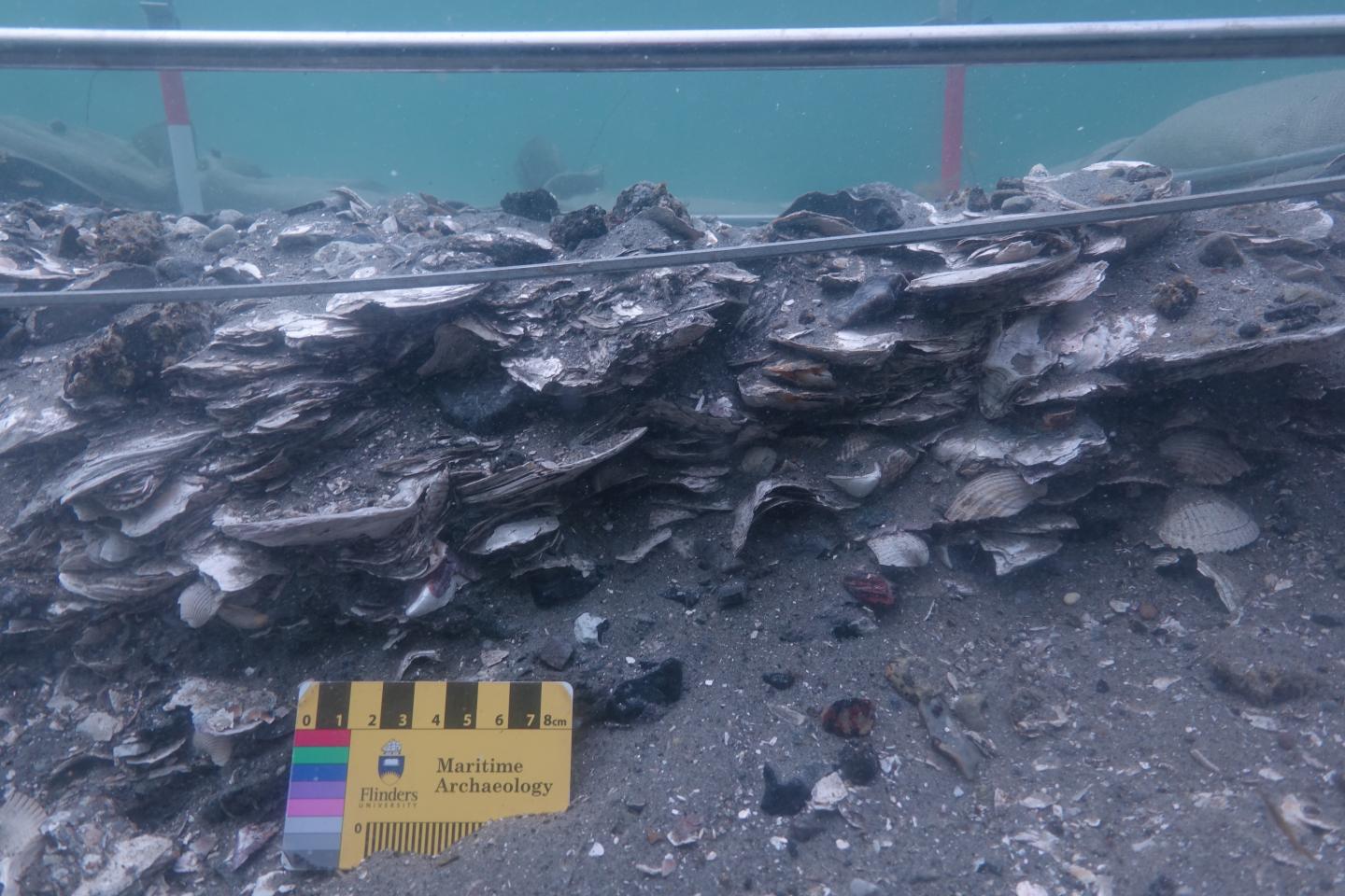
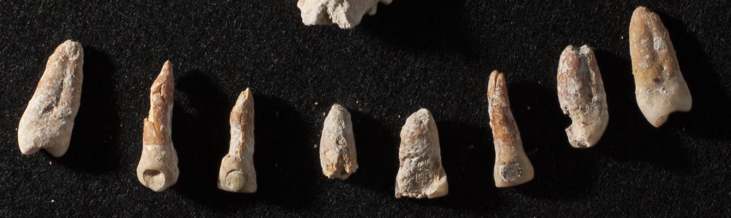

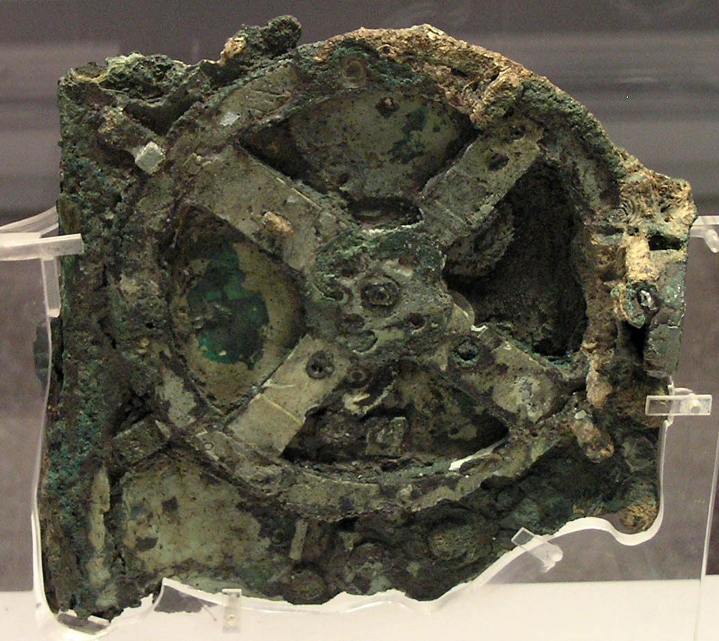


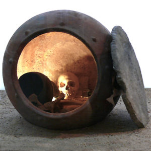
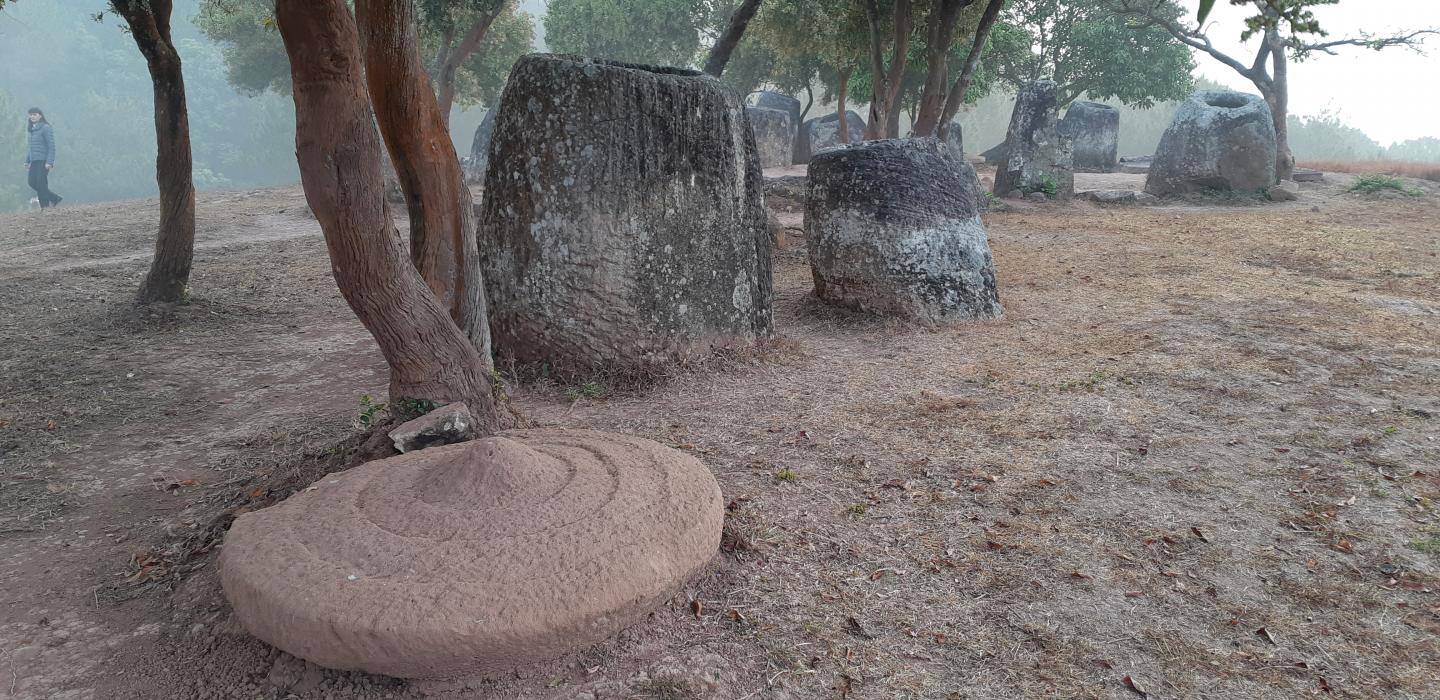
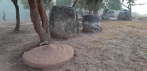
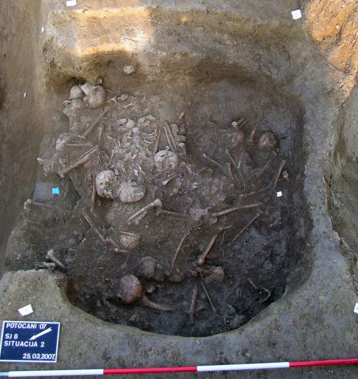
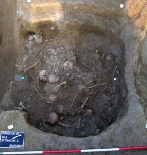
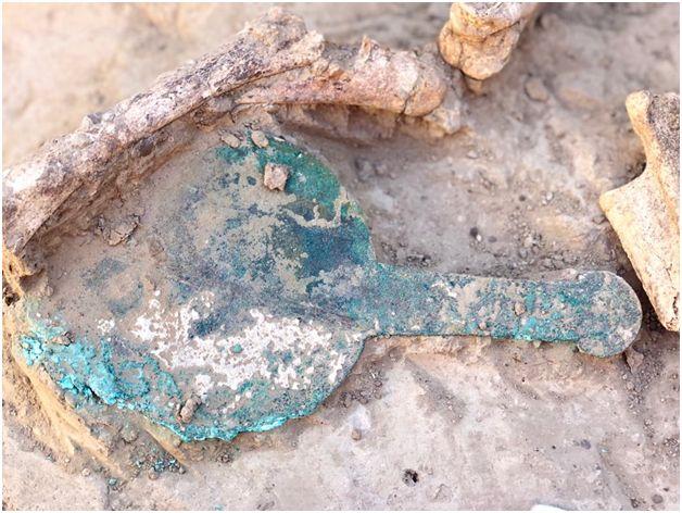

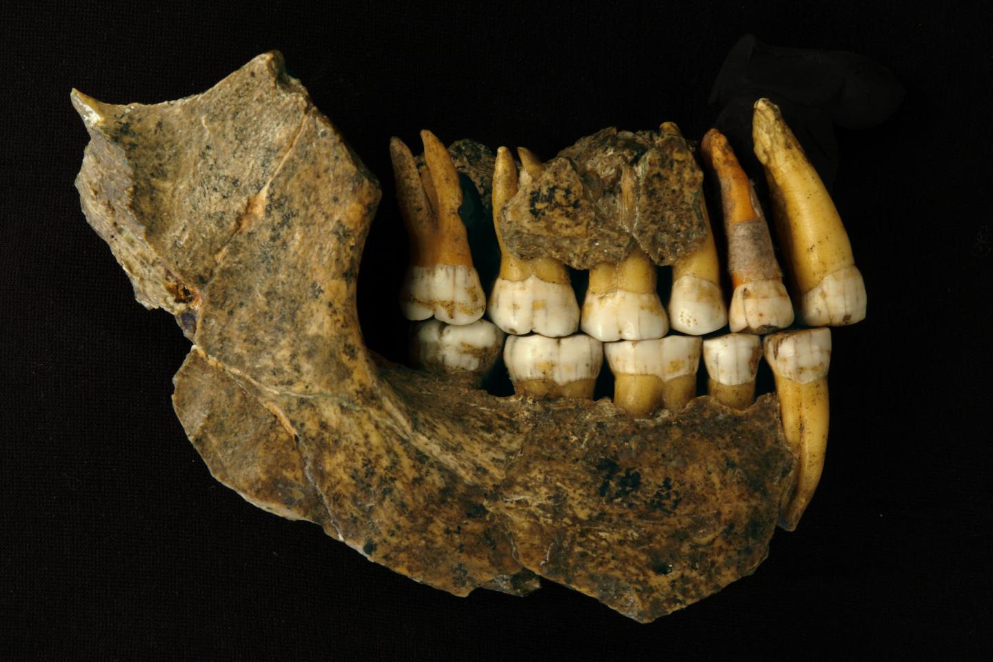

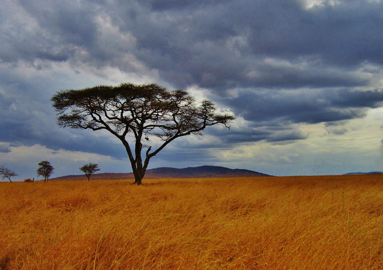


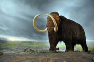
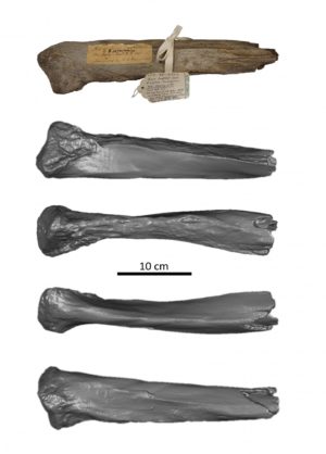
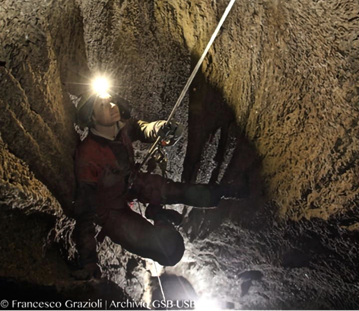
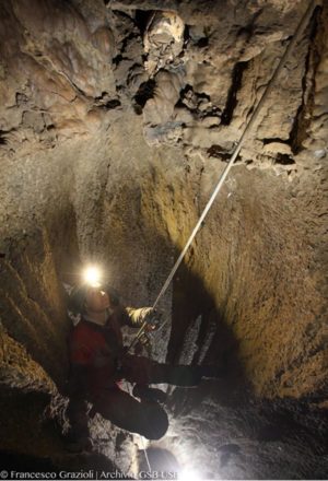
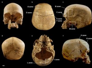
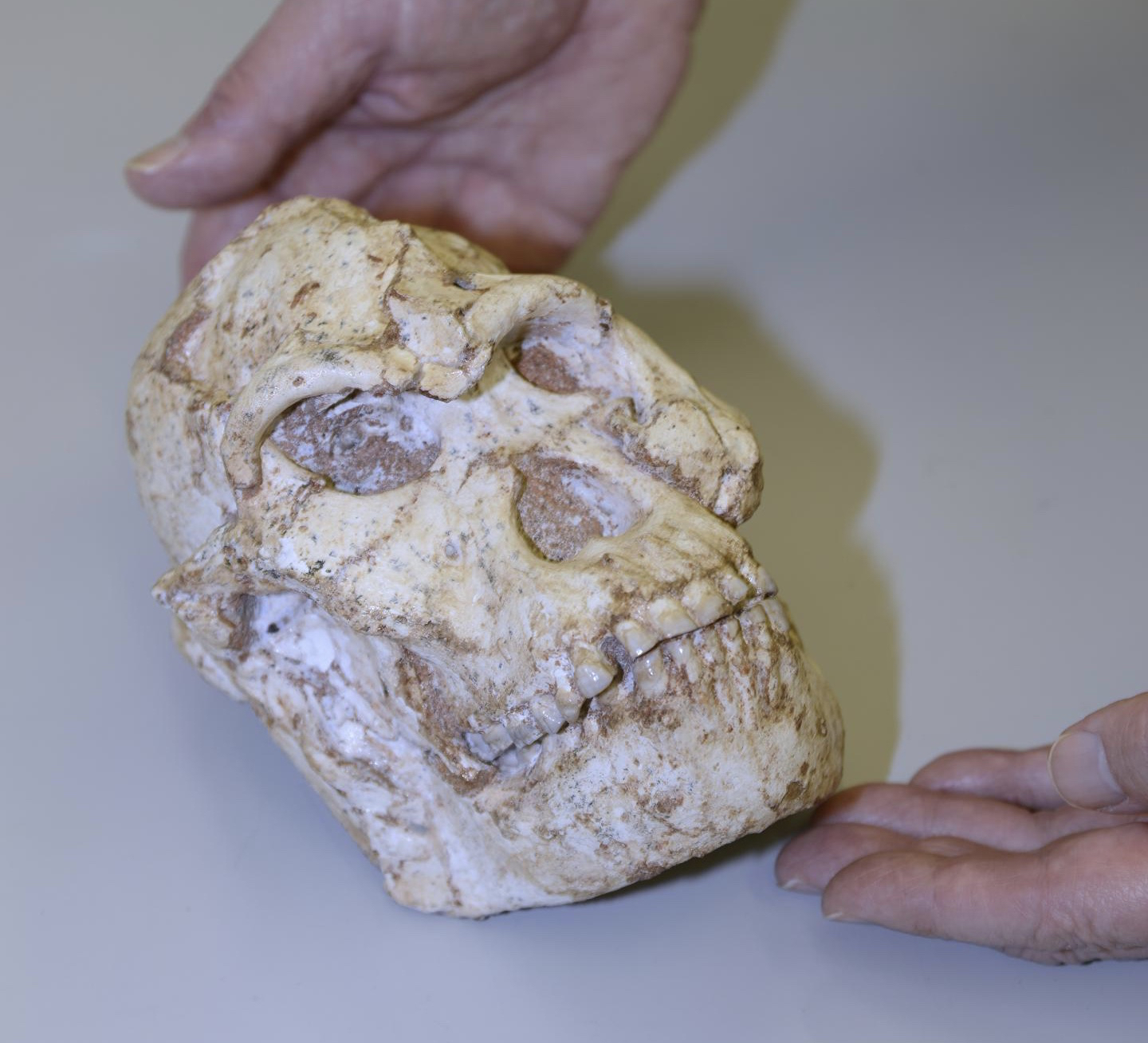

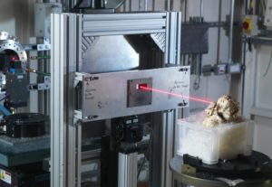
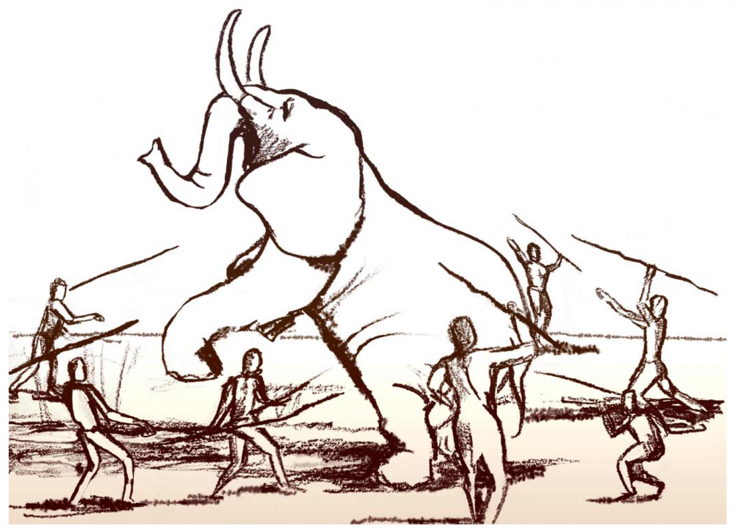
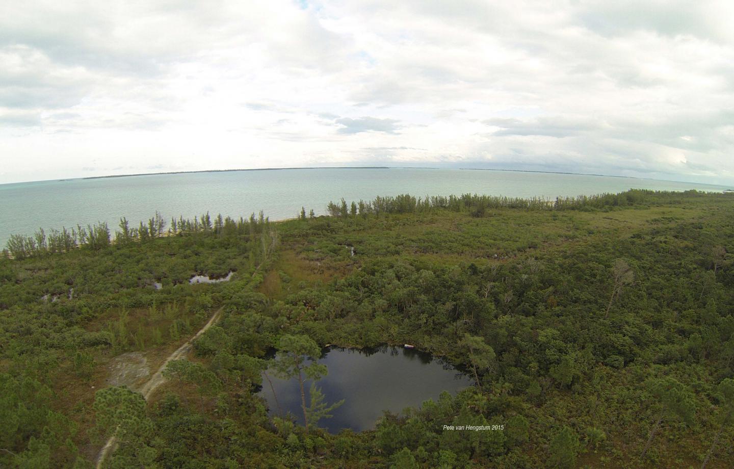

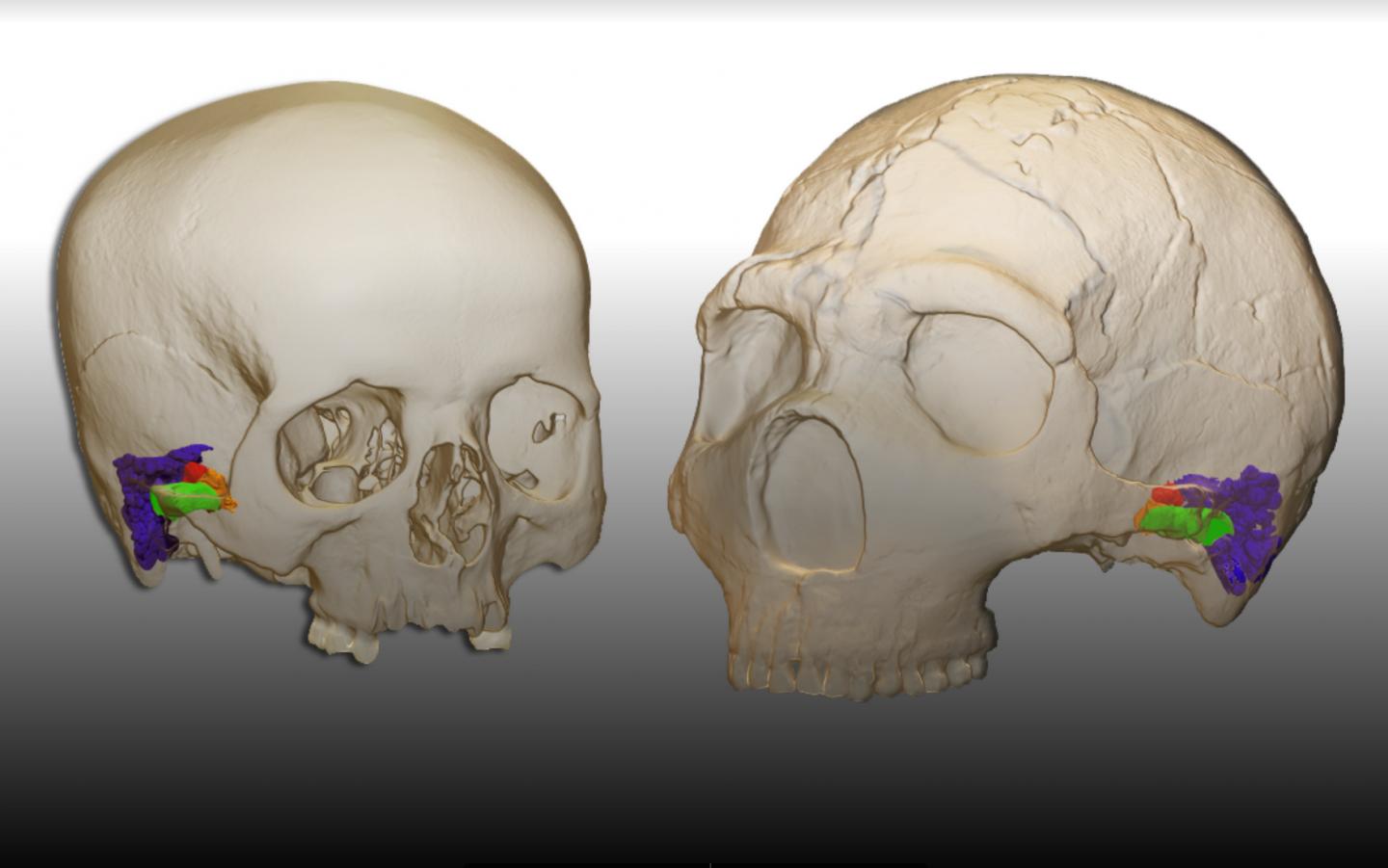
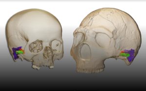

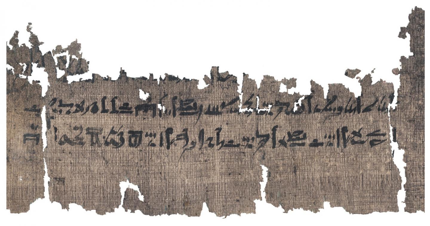
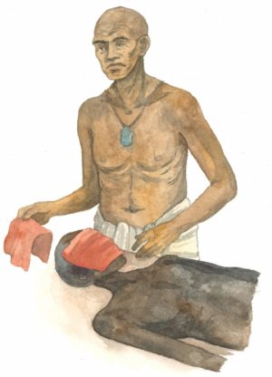
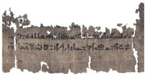

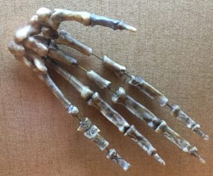
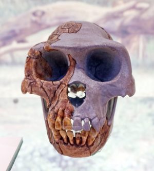
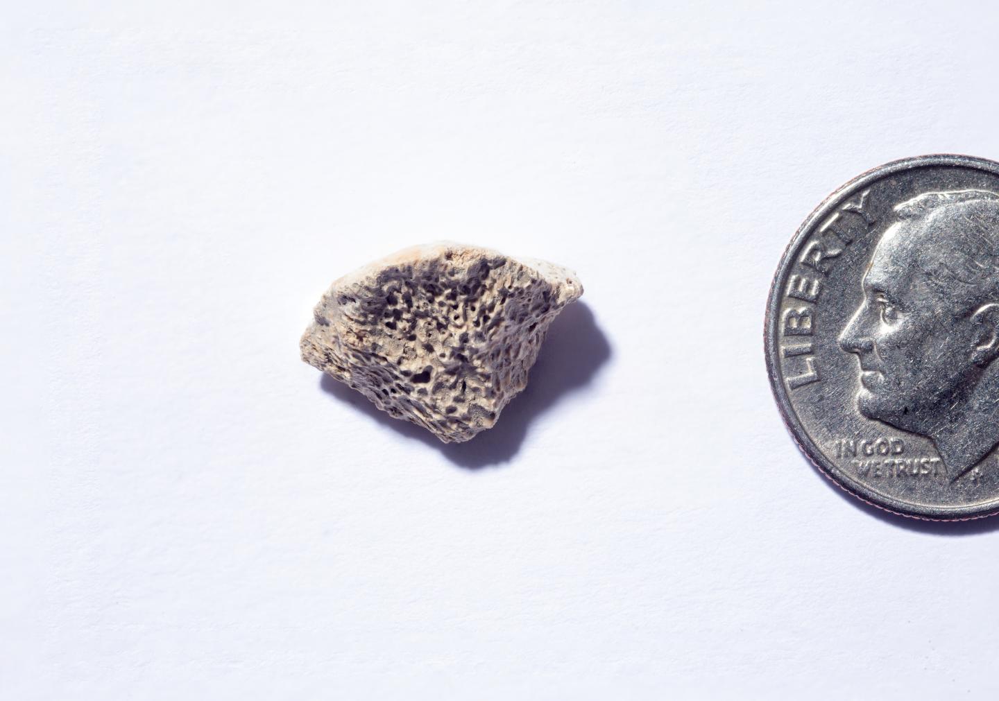

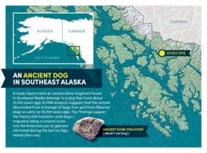


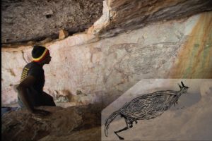

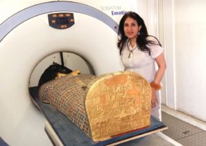

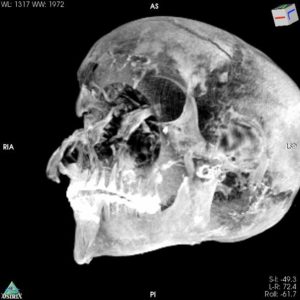


